Hepatosplenic T cell lymphoma with no expression of cytotoxic molecules
- PMID: 12890821
- PMCID: PMC1770035
- DOI: 10.1136/jcp.56.8.631
Hepatosplenic T cell lymphoma with no expression of cytotoxic molecules
Abstract
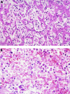
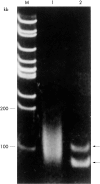
Hepatosplenic T cell lymphoma is defined as an extranodal and systemic neoplasm derived from cytotoxic T cells. This report describes a postmortem case of T cell lymphoma that showed histological features of hepatosplenic T cell lymphoma but did not express cytotoxic molecules. The patient was a 57 year old man who presented with severe icterus and hepatosplenomegaly, followed by an aggressive clinical course. The liver and spleen were enlarged, weighing 2000 g and 360 g, respectively. Histologically, the liver, spleen, and bone marrow were entirely affected by lymphoma, comprising pleomorphic small and large cells, which displayed sinusoidal infiltration in the liver, diffuse infiltration in the splenic cord, and interstitial/diffuse infiltration with fibrosis in the bone marrow. Lymphoma cells showed positivity for CD3 epsilon, CD8, and CD45RO and clonal rearrangement of the TCRgamma gene by the polymerase chain reaction on paraffin wax embedded sections. However, they were negative for TIA-1 and granzyme B, in addition to betaF1, CD4, and CD56. Few neoplastic cells were stained for Epstein-Barr virus encoded mRNA 1. These findings indicate that this case might represent a variant of hepatosplenic T cell lymphoma despite the absence of cytotoxic molecules.
Figures


References
-
- Farcet JP, Gaulard P, Marolleau JP, et al. Hepatosplenic T-cell lymphoma: sinusal/sinusoidal localization of malignant cells expressing the T-cell receptor gamma delta. Blood 1990;75:2213–19. - PubMed
-
- Jaffe ES, Ralfkiaer E. Hepatosplenic T-cell lymphoma. In: Jaffe ES, Harris NL, Stein H, et al, eds. World Health Organization classification of tumors. Pathology and genetics of tumors of haematopoietic and lymphoid tissues. Lyon: IARC Press, 2001:210–11.
-
- Macon WR, Levy NB, Kurtin PJ, et al. Hepatosplenic αβT-cell lymphomas. Report of 14 cases and comparison with hepatosplenic γδT-cell lymphomas. Am J Surg Pathol 2001;25:285–96. - PubMed
-
- Anderson P, Nagler-Anderson C, O’Brien C, et al. A monoclonal antibody reactive with a 15-kDa cytoplasmic granule-associated protein defines a subpopulation of CD8+ T lymphocytes. J Immunol 1990;144:574–82. - PubMed
-
- Cooke CB, Krenacs L, Stetler-Stevenson M, et al. Hepatosplenic T-cell lymphoma: a distinct clinicopathologic entity of cytotoxic gamma delta T-cell origin. Blood 1996;88:4265–74. - PubMed
Publication types
MeSH terms
Substances
LinkOut - more resources
Full Text Sources
Medical
Research Materials
Miscellaneous
