Review
. 2003 Jul;203(1):1-19.
doi: 10.1046/j.1469-7580.2003.00203.x.

Neurotrophins and the immune system

Affiliations
Review

Neurotrophins and the immune system

José A Vega et al. J Anat. 2003 Jul.

Abstract

The neurotrophins are a family of polypeptide growth factors that are essential for the development and maintenance of the vertebrate nervous system. In recent years, data have emerged indicating that neurotrophins could have a broader role than their name might suggest. In particular, the putative role of NGF and its receptor TrkA in immune system homeostasis has become a much studied topic, whereas information on the other neurotrophins is scarce in this regard. This paper reviews what is known about the expression and possible functions of neurotrophins and their receptors in different immune tissues and cells, as well as recent data obtained from studies of transgenic mice in our laboratory. Results from studies to date support the idea that neurotrophins may regulate some immune functions. They also play an important role in the development of the thymus and in the survival of thymocytes.


Figures

Fig. 1
Fig. 1
Schematic representation of the structure of Trk and p75NTR neurotrophin receptors, and of the neurotrophins that bind each of them: thick arrows represent the primary receptor–ligand pairing; thin arrows indicate other binding possibilities. Two different isoforms of TrkA have been isolated, and termed TrkAI and TrkAII. Three TrkAI isoforms are known (shown at the left of the figure). In the thymus, two specific TrkAI isoforms have been isolated (arrows at bottom left), which show total or partial deletion of amino cysteine-rich regions of the extracellular domain. TrkAII differs from TrkAI in that it carries a small insertion of six amino acids next to the transmembrane domain (hexagon). With regard to TrkB and TrkC, full-length (FL), tyrosine kinase truncated (TK-T1 and TK-T2), and tyrosine kinase inserted (14, 25 and 39, equivalent to the number of amino acids forming the insertion) isoforms have been isolated. NH2 CRR, amino cysteine-rich regions; COOH-CRR, carboxy cysteine-rich regions; CRD, cysteine-rich domains; LRR, leukine-rich region; TK, tyrosine kinase domain.
Fig. 2
Fig. 2
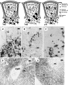
The upper pictures illustrate the localization of TrkA-(left), TrkB-(middle) and p75NTR (right)-positive cells in the rodent thymus; receptor-expressing cells are represented in black. Below are shown the corresponding TrkA (A), TrkB (B) and p75NTR (C) immunostained sections. The cells expressing TrkA are subcapsular and medullar thymic epithelial cells (mouse); those showing TrkB immunoreactivity, concentrated at the cortico-medullary border (arrows), are macrophages (rat), and p75NTR immunoreactivity is confined to a subpopulation of medullar thymic epithelial cells (arrows, rat). The lower images correspond to morphological aspects of functionally trkA- and trkB-deficient mice. The trkA-kinase –/– mouse thymus (D) is characterized by disorganization of the thymic architecture and the presence of medullar endodermic cysts containing amorphous material and cell debris, whereas the trkB-kinase –/– mouse (E) shows images of apoptotic lymphocyte death, especially in the cortex. c, cortical; m, medullar. Scale bar = 5 µm.
Fig. 3
Fig. 3
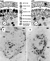
The upper pictures illustrate the localization of TrkB-(left) and p75NTR (right)-positive cells in the rat spleen; receptor-expressing cells are represented in black. Below are shown the corresponding TrkB (A) and p75NTR (B) immunostained sections. TrkB-immunoreactive cells include MMM and scattered white pulp macrophages, whereas p75NTR-positive cells are a subpopulation of dendritic cells. PALS, periarteriolar lymphatic sheath; F, follicle; MZ, marginal zone; CA, central arteriole; MM, marginal metallophilic. Scale bar = 5 µm.
Fig. 4
Fig. 4
Schematic representation of the possible sources and targets of NGF and BDNF in the mammalian thymus.

Similar articles

Cited by

References

    1. Abramchik SS, Yermakova VN, Kaliunov RM, Tanina RM, Tumilovich MK. The immunomodulatory effect of nerve growth factor. J. Neurosci. Res. 1988;19:349–356. - PubMed
    1. Aloe L, Levi-Montalcini R. Mast cell increase in tissues of neonatal rats injected with the nerve growth factor. Brain Res. 1977;133:358–366. - PubMed
    1. Aloe L. The effect of nerve growth factor and its antibody on mast cells in vivo. J. Neuronimmunol. 1988;18:1–12. - PubMed
    1. Aloe L, De Simone R. NGF primed spleen cells injected in brain of developing rats differentiate into mast cells. Int. J. Dev. Neurosci. 1989;7:565–573. - PubMed
    1. Aloe L, Tuveri MA, Carcassi U, Levi-Montalcini R. Nerve growth factor in the synovial fluid of patients with chronic arthritis. Arthritis Rheumatism. 1992;35:351–355. - PubMed

Publication types

MeSH terms