. 2003 Jul;203(1):31-56.
doi: 10.1046/j.1469-7580.2003.00208.x.

Ultrastructural distribution of glycinergic and GABAergic neurons and axon terminals in the rat dorsal cochlear nucleus, with emphasis on granule cell areas

Affiliations

Ultrastructural distribution of glycinergic and GABAergic neurons and axon terminals in the rat dorsal cochlear nucleus, with emphasis on granule cell areas

Lorenzo Alibardi. J Anat. 2003 Jul.

Abstract

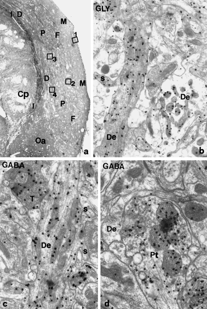
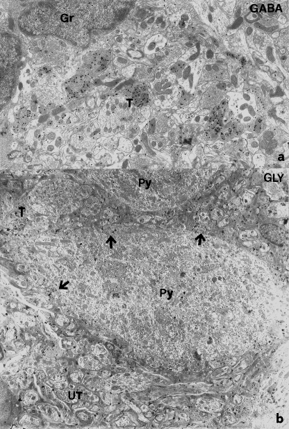
A knowledge of neurotransmitters in the neurons of the rat cochlear nuclear complex is of importance in understanding the function of auditory circuits. Using post-embedding ultrastructural immunogold labelling, the distribution of glycinergic and GABAergic neurons and axonal terminals has been studied in the molecular, fusiform and polymorphic layers of the rat dorsal cochlear nucleus (DCN). This technique is not limited by the penetration of antibodies into the nervous tissue as in pre-embedding methods, and allows a fine neurochemical mapping of the nervous tissue. Numerous glycinergic and GABAergic axon terminals contain pleomorphic and flat synaptic vesicles, and are present in all layers (1, 2, 3) of the dorsal cochlear nucleus. Glycine and GABA-negative large terminals (mossy fibres) are mainly seen in granule cell areas of layer 2 (fusiform layer). Mossy fibres contact the dendrites of GABA- and glycine-negative granule cells and of the few unipolar brush cells (excitatory neurons). The least common cells in the granule cell areas are GABAergic and glycinergic Golgi-stellate neurons. In unipolar brush cells, aggregations of vesicles seem to be the origin of their characteristic ringlet-bodies. Golgi-stellate cells send their inhibitory terminals to the dendrites of granule and unipolar brush cells, occasionally directly to mossy fibres. Small or (less frequently) large GABAergic terminals contact the soma or the main dendrite of unipolar brush cells. The circuit of a hypothetical functional unit of neurons in the DCN is proposed. The inputs from auditory tonotopic or non-auditory non-tonotopic mossy fibres eventually reach pyramidal cells through axons from the granule cells or unipolar brush cells. Pyramidal cells convey an excitatory signal from the DCN to higher mesencephalic nuclei for further elaboration of the acoustic signal.


Figures

Fig. 1
Fig. 1
(a) Light microscopic view of rat DCN with the layer subdivisions. Oa, octopus cell area. Cp, inferior cerebellar peduncle. The squares are the corresponding areas of the molecular layer (1), fusiform layer (2), polymorphic layer (3) and polymorphic layer near dorsal acoustic stria (4), which have been analysed under the electron microscope for immunolabelling background (see following figures). Toluidine blue. ×70. (b,c) Immunolabelling in the molecular layer, localized as in square 1 in (a). (b) Glycine labelling of electron-dense and pale dendrites among unlabelled neuropile. ×18 700. (c) GABA labelling of pale dendrite contacted by a labelled P-terminal. ×25 000. (d) GABAergic P-terminal contacting immunonegative dendrites. ×30 000.
Fig. 2
Fig. 2
(a) Superficial molecular layer (position similar to square 1 in Fig. 1a) under the ependymal epithelium. Some intensely GABA-labelled boutons are seen among the immunonegative neuropil. ×6400. (b) Lower molecular layer showing many GABA-labelled myelinated axons and terminals among an immunonegative neuropil. ×8000.
Fig. 3
Fig. 3
(a) Superficial molecular layer beneath the ependyma (see square 1 of Fig. 1) where numerous terminals are more intensely labelled than the background. ×6600. (b) Detail of the molecular layer illustrating the diffuse background glycine immunolabelling among intensely positive terminals. ×9800.
Fig. 4
Fig. 4
(a) GABA-immunonegative granule cells in neuropil of upper layer 2 (corresponding to square 2 of Fig. 1) where numerous GABA-positive terminals are seen. ×10 600. (b) Glycine-immunonegative pyramidal cells in layer 2 (see corresponding position in square 2 of Fig. 1) surrounded by many labelled axo-somatic boutons (arrows). Numerous labelled terminals are seen among unlabelled myelinated axons and neuropil structures. ×5000.
Fig. 5
Fig. 5
(a) GABA-positive Golgi-stellate cell in layer 2 associated with an immunonegative granule cell within a cluster of small cells. The arrow indicates immunonegative axo-somatic R-boutons. ×12 700. (b) Granule-like cell in layer 2 showing the perinuclear chromatin and a ringlet-body (inset at ×34 200). ×17 200. (c) Detail of microtubules close to a ringlet-body within a cell elongation. ×52 500.
Fig. 6
Fig. 6
(a) Intensely glycine-labelled cartwheel neuron, including the round nucleus and dendrite, within a diffusely labelled neuropil in upper layer 2. ×6000. (b) GABA-labelled Golgi-stellate neuron among an immunegative neuropil in upper layer 2. ×14 700.
Fig. 7
Fig. 7
(a) Small, pale Golgi-stellate cell with perinuclear chromatin labelled for GABA in layer 2 (compare the lack of labelling in the nearby pyramidal cell). ×14 700. (b) Intensely GABA-labelled Golgi-stellate cell with indented nucleus in polymorphic layer. ×15 900. GABA-labelled boutons rich in pleomorphic vesicles (left inset, ×30 000). Glycine-labelled bouton rich in pleomorphic and flat vesicles (right inset, ×30 000).
Fig. 8
Fig. 8
(a) Glycine-negative unipolar brush cell in layer 2 with euchromatic nucleus, Golgi apparatus, ringlet-body and dendrioles (arrowheads). Arrows indicate two glycine-positive boutons. ×14 000. (b) Detail of the ringlet-body in (a). The arrow indicates a cytoplasmic bleb contacting a glycine-positive FP-bouton. ×28 500. (c) Continuity between the Golgi apparatus and ringlet-body of a unipolar brush cell. Coated vesicles blebbing from dictyosomes (arrows) and a dense-cored vesicle (arrowhead) are seen. ×56 000.
Fig. 9
Fig. 9
(a) Ringlet body of a unipolar brush cell. Continuity between Golgi vesicles and round vesicles (arrowheads), and with open vesicles forming ringlets (arrows) within amorphous material. ×45 600. (b) Detail of blebbing-coated vesicles (arrowheads) and round vesicles (arrow) in Golgi apparatus of unipolar brush cell. ×44 500. (c) Glomerulus close to the cell body of a unipolar brush cell with indented nucleus. A mossy fibre directly contacts the brush cell and is surrounded by sectioned dendrites of a unipolar brush cell. The dendrites are externally contacted by GABA-positive boutons (arrowheads), one of which also directly reaches the cell body of the brush cell (double arrowhead). ×10 400.
Fig. 10
Fig. 10
(a,b) Glomeruli in layer 2 localized distally from unipolar brush cells. (a) GABA-negative mossy fibre surrounded by long-sectioned pale dendrites of unipolar brush cell, which are externally contacted by GABA-positive boutons (asterisks). ×16 500. (b) Glycine-negative mossy fibre surrounded by some unipolar brush cell dendrites, externally contacted by glycine-positive terminals (asterisks). The presence of two blebs (arrowheads) suggests that this mossy fibre may derive from a unipolar brush cell. ×12 800.
Fig. 11
Fig. 11
(a) Large GABA-positive pleomorphic bouton forming symmetric synapses (arrowheads) and a slightly asymmetric synapse (arrow) with unipolar cell dendrites in layer 2. ×25 000. (b) Undulatory-shaped, large GABA-positive synaptic pleomorphic terminal in layer 2 forming asymmetric synapses (arrows) to a GABA-negative post-synaptic unipolar brush cell large dendrite. ×34 700. (c) Glycine-positive undulatory-shaped P-terminal making a symmetric synapse (arrow) with a dendrite of unipolar brush cell. ×33 000.
Fig. 12
Fig. 12
(a) GABA-positive flat Golgi-stellate cell with indented nucleus and opposite dendrites (arrows) in lower layer 3 (corresponding to square 4, Fig. 1). ×8300. (b) GABA-labelled P-boutons forming symmetric synapses (arrow) onto the initial dendrite of unipolar brush cell in layer 2. ×30 300. (c) Large GABA terminal with pleomorphic vesicles surrounding small unipolar brush or granule cell distal dendrite in layer 2. ×39 200.
Fig. 13
Fig. 13
(a) GABA-negative mossy fibre contacted by two GABAergic P-terminals, one of which forms a synapse (arrow). A terminal containing small round vesicles is also part of this synaptic structure. ×25 700. (b) GABA-negative cell in the polymorphic layer (see square 4, Fig. 1) receiving two immunolabelled axo-somatic terminals (arrows). Among myelinated axons GABAergic terminals are seen. ×4800.
Fig. 14
Fig. 14
(a) Glycine-negative large-sized giant neuron in layer 3 (see square 3, Fig. 1) with some immunopositive axo-somatic boutons (arrows). Immunolabelled terminals are seen among unlabelled axons and terminals. ×4800. (b) Glycinergic vertical cell in lowermost layer 3 (see square 4 of Fig. 1) contacted by two immunopositive axo-somatic boutons (arrows) and one immunonegative bouton (arrowhead). An immunonegative granule–unipolar brush-like cell is also reached by two glycinergic boutons (arrows). Numerous surrounding myelinated axons are labelled. ×6300.
Fig. 15
Fig. 15
(a,b) Parallel section of the same vertical neuron in layer 3 (as in square 3, Fig. 1). The different arrows indicate the same terminals as they appear after glycine or GABA immunolabelling. (a) A small quantity of glycine-positive (arrow and double arrow) and glycine-negative (arrowhead) boutons contact this glycine-positive cell. ×8200. (b) Parallel section of the same cell showing that it is GABA-negative. One of the same boutons (arrow) co-localizes GABA, another (double arrowhead) is GABA-negative and the intermediate bouton (arrowhead) is only GABA-positive. ×8200.
Fig. 16
Fig. 16
(a) Glycine-negative unipolar brush cell with the dendrite close to a glycine-positive, possibly vertical neuron in lower layer 3 (see square 4, Fig. 1). The square indicates the areas shown at higher magnification in (b). Surrounding boutons are immunonegative (arrows) or glycine-positive (asterisks). ×8000. (b) Area shown in the square in (a), showing the detail of a ringlet-body (arrowhead) in continuity with vesicles (arrow) of the Golgi apparatus. ×44 500. (c) Ringlet-body in distal unipolar cell dendrite in layer 3. ×20 000.
Fig. 17
Fig. 17
(a) Large GABA-labelled P-bouton in layer 3 (corresponding to square 3 of Fig. 1) apposed to a mossy fibre (arrows). ×31 000. (b) Mossy fibre contacting (arrow) a large GABA-labelled P-terminal among heavily myelinated axons in layer 3. ×22 500.
Fig. 18
Fig. 18
Schematic drawing of two possible units within granule cell areas. Circuit I is centred on a unipolar brush cell whereas circuit II is centred on granule cells. A mixed unit I and II may also be possible. Ax, axon; De, dendrite; G, granule cell with apical T branching axons to originate parallel fibres; GS, Golgi-stellate cell; eMF, extrinsic mossy fibre; iMF, intrinsic mossy fibre; + or – indicate an excitatory or inhibitory stimulus. U, unipolar brush cell. The arrows indicate the direction of the input. The arrowheads indicate a direct inhibitory (GABA/glycinergic) terminal onto the mossy fibre.

Similar articles

Cited by

References

    1. Alibardi L. Characterization of tuberculo-ventral neurons in dorsal cochlear nucleus of the guinea pig. J. Submicrosc. Cytol. Pathol. 1999a;31:295–300. 10.1046/j.1469-7580.2003.00208.x. - DOI - PubMed
    1. Alibardi L. Fine structure, synaptology and immunocytochemistry of large neurons in the rat dorsal cochlear nucleus connected to the inferior colliculus. J. Brain Res. 1999b;39:427–437. 10.1046/j.1469-7580.2003.00208.x. - DOI - PubMed
    1. Alibardi L. Identification of tuberculoventral neurons in the polymorphic layer of the rat dorsal cochlear nucleus. Eur. J. Morphol. 2000a;38:153–166. 10.1046/j.1469-7580.2003.00208.x. - DOI - PubMed
    1. Alibardi L. Cytology of large neurons in the guinea pig dorsal cochlear nucleus. Eur. J. Histochem. 2000b;44:365–375. 10.1046/j.1469-7580.2003.00208.x. - DOI - PubMed
    1. Alibardi L. Cytology, synaptology and immunocytochemistry of commissural neurons and their putative axonal terminals in the dorsal cochlear nucleus of the rat. Ann. Anat. 2000c;182:207–220. 10.1046/j.1469-7580.2003.00208.x. - DOI - PubMed

LinkOut - more resources