. 2003 Jul;203(1):89-99.
doi: 10.1046/j.1469-7580.2003.00195.x.

Muscle satellite (stem) cell activation during local tissue injury and repair

Affiliations

Muscle satellite (stem) cell activation during local tissue injury and repair

Maria Hill et al. J Anat. 2003 Jul.

Abstract

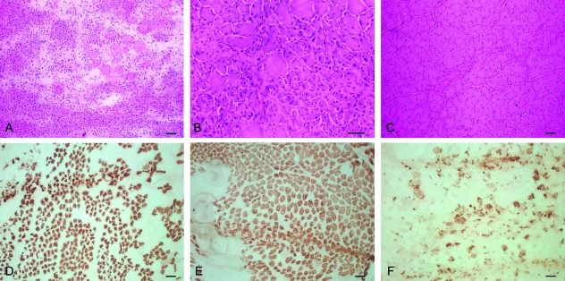
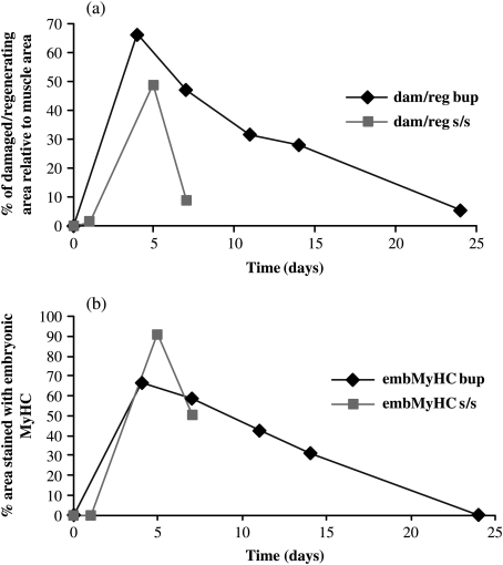
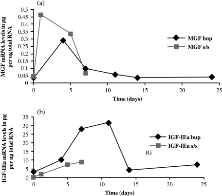
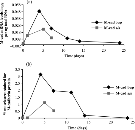
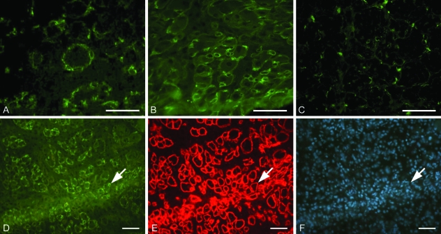
In post-mitotic tissues, damaged cells are not replaced by new cells and hence effective local tissue repair mechanisms are required. In skeletal muscle, which is a syncytium, additional nuclei are obtained from muscle satellite (stem) cells that multiply and then fuse with the damaged fibres. Although insulin-like growth factor-I (IGF-l) had been previously implicated, it is now clear that muscle expresses at least two splice variants of the IGF-I gene: a mechanosensitive, autocrine, growth factor (MGF) and one that is similar to the liver type (IGF-IEa). To investigate this activation mechanism, local damage was induced by stretch combined with electrical stimulation or injection of bupivacaine in the rat anterior tibialis muscle and the time course of regeneration followed morphologically. Satellite cell activation was studied by the distribution and levels of expression of M-cadherin (M-cad) and related to the expression of the two forms of IGF-I. It was found that the following local damage MGF expression preceded that of M-cad whereas IGF-IEa peaked later than M-cad. The evidence suggests therefore that an initial pulse of MGF expression following damage is what activates the satellite cells and that this is followed by the later expression of IGF-IEa to maintain protein synthesis to complete the repair.


Figures

Fig. 1
Fig. 1
Transverse sections of rat TA muscle stained with haematoxylin and eosin demonstrating maximal damage at (a) 4 days after bupivacaine injection, (b) 5 days following stretch and stimulation and (c) recovery at 14 days following bupivacaine injection where central nuclei were present in the regenerated fibres. Bupivacaine injection caused massive muscle fibre degeneration as seen in (a) with a small number of fibres in the periphery of the section that survived the bupivacaine insult. (d–f) Transverse sections of the same TA muscles as above, stained with embryonic myosin heavy chain to determine the regenerating muscle fibres at each time point following both damage models. At both 4 and 5 days following bupivacaine injection and stretch and stimulation, embryonic myosin heavy chain stained heavily whereas at 14 days only a few regenerating muscle fibres were present (f). Scale bar = 50 µm.
Fig. 2
Fig. 2
(a) Mean percentage of damaged–regenerating muscle fibre area in relation to the whole muscle section in both damage models. There is a continuing decrease in the damaged–regenerating area after 4 days following bupivacaine injection and 5 days after stretch and stimulation where maximal damage was present in both. (b) The same pattern was seen with the embryonic myosin heavy chain staining the regenerating area in both models. Two-way analysis of variance (anova) was used to determine significant differences among the means. N = 4 for bupivacaine model and N = 6 for stretch/stimulation model for each time point. Experimental muscles were compared with those of the untreated animals at zero time and all differences were significant at P < 0.01 up to 15 days post-injury.
Fig. 3
Fig. 3
mRNA levels of MGF and IGF-IEa isoforms in the two models of muscle damage. MGF was maximally expressed as early as 1 day following stretch and stimulation and 4 days following bupivacaine injection (a), whereas IGF-IEa was maximally expressed later at 7 and 11 days following injury (b). Two-way analysis of variance (anova) was used to determine significant differences among the means. N = 6 for stretch/stimulation model and N = 4 for bupivacaine model for each time point. MGF measurements were significant at P < 0.01 for up to 5 days and IGF-IEa for up to 11 days.
Fig. 4
Fig. 4
M-cad mRNA and protein expression in damaged TA muscle following stretch and stimulation and bupivacaine injection. M-cad mRNA levels (a) seemed to peak at 4–5 days after bupivacaine injection after stretch and stimulation and (b) M-cad protein levels peak shortly after but rapidly decrease once regeneration has commenced. All measurements of M-cad mRNA and protein were significantly different between the experimental muscles up to 11 days compared with normal resting values.
Fig. 5
Fig. 5
M-cad staining in a cross-section of TA muscle (a) 4 days after bupivacaine injection, (b) 5 days following stretch and stimulation and (c) 14 days following bupivacaine injection. In order to verify the identity of the M-cad-positive cells as satellite cells in (d), triple staining with laminin antibody (e) to identify the basal lamina and bis-benzimide (f) for nuclei staining was performed in order to check that M-cad was staining the satellite cells underneath the basal lamina. M-cad staining (arrows in d–f) was seen in the form of positive rings at the periphery of the muscle fibres but underneath the basal lamina which was much more obvious in the fibres of the damaged muscles (d). Scale bar = 50 µm.

Similar articles

Cited by

References

    1. Adams GR. Role of insulin-like growth factor-I in the regulation of skeletal muscle adaptation to increased loading. Exerc. Sport Sci. Rev. 1998;26:31–60. - PubMed
    1. Adams GR. Exercise effects on muscle insulin signalling and action. Invited Review: Autocrine/paracrine IGF-I and skeletal muscle adaptation. J. Appl. Physiol. 2002;93:1159–1167. - PubMed
    1. Aziz-Ullah, Goldspink G. Distribution of mitotic nuclei in the biceps brachii of the mouse during post-natal growth. Anat. Rec. 1974;179:115–118. - PubMed
    1. Beauchamp JR, Heslop L, Yu DS, Tajbakhsh S, Kelly RG, Wernig A, et al. Expression of CD34 and Myf5 defines the majority of quiescent adult skeletal muscle satellite cells. J. Cell Biol. 2000;151:1221–1234. - PMC - PubMed
    1. Bornemann A, Schmalbruch H. Immunocytochemistry of M-cadherin in mature and regenerating rat muscle. Anat. Rec. 1994;239:119–125. - PubMed

Publication types

MeSH terms

LinkOut - more resources