. 2003 Jul 26;362(9380):263-70.
doi: 10.1016/S0140-6736(03)13967-0.

Newly discovered coronavirus as the primary cause of severe acute respiratory syndrome

Affiliations

Newly discovered coronavirus as the primary cause of severe acute respiratory syndrome

Thijs Kuiken et al. Lancet. .

Abstract

Background: The worldwide outbreak of severe acute respiratory syndrome (SARS) is associated with a newly discovered coronavirus, SARS-associated coronavirus (SARS-CoV). We did clinical and experimental studies to assess the role of this virus in the cause of SARS.

Methods: We tested clinical and postmortem samples from 436 SARS patients in six countries for infection with SARS-CoV, human metapneumovirus, and other respiratory pathogens. We infected four cynomolgus macaques (Macaca fascicularis) with SARS-CoV in an attempt to replicate SARS and did necropsies on day 6 after infection.

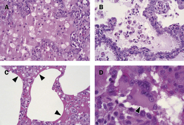
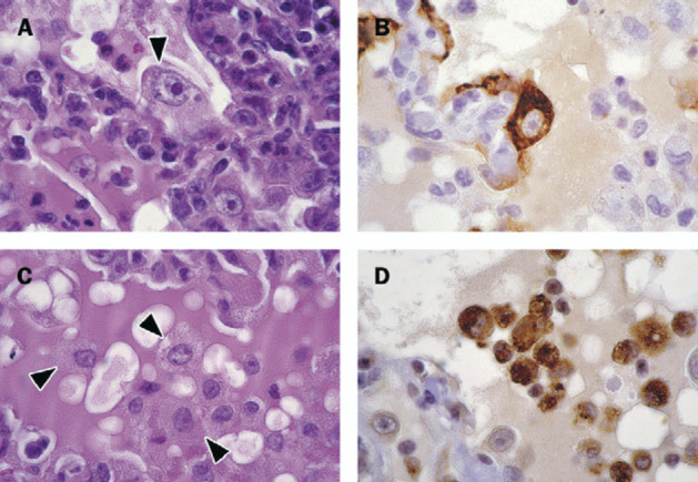
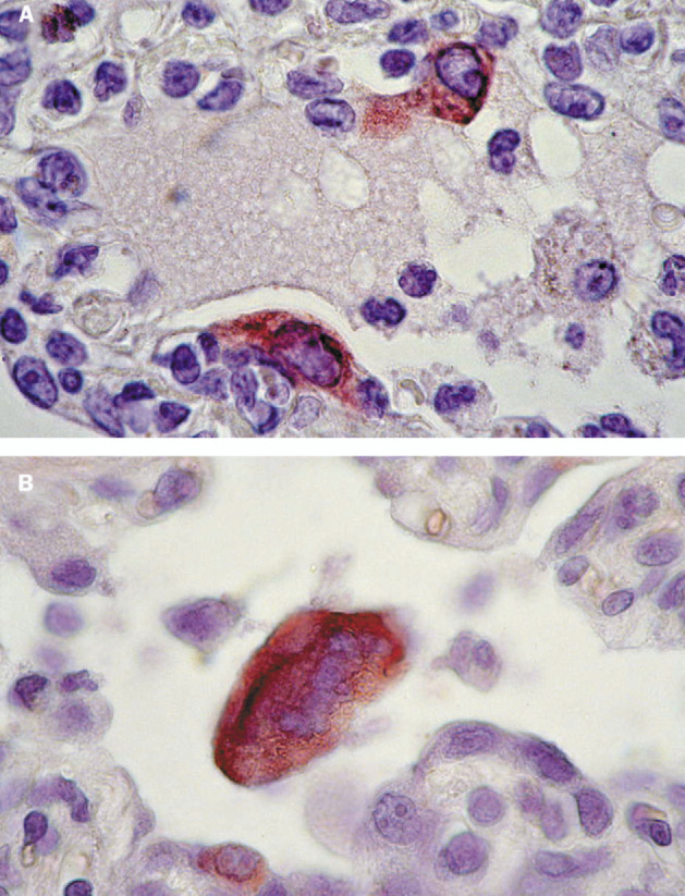
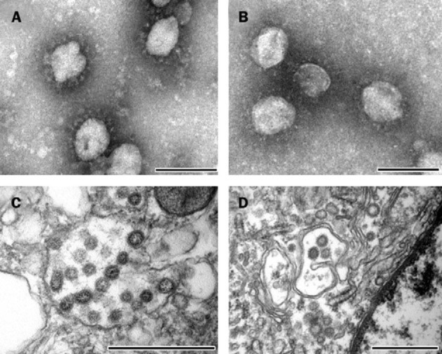
Findings: SARS-CoV infection was diagnosed in 329 (75%) of 436 patients fitting the case definition of SARS; human metapneumovirus was diagnosed in 41 (12%) of 335, and other respiratory pathogens were diagnosed only sporadically. SARS-CoV was, therefore, the most likely causal agent of SARS. The four SARS-CoV-infected macaques excreted SARS-CoV from nose, mouth, and pharynx from 2 days after infection. Three of four macaques developed diffuse alveolar damage, similar to that in SARS patients, and characterised by epithelial necrosis, serosanguineous exudate, formation of hyaline membranes, type 2 pneumocyte hyperplasia, and the presence of syncytia. SARS-CoV was detected in pneumonic areas by virus isolation and RT-PCR, and was localised to alveolar epithelial cells and syncytia by immunohistochemistry and transmission electron microscopy.

Interpretation: Replication in SARS-CoV-infected macaques of pneumonia similar to that in human beings with SARS, combined with the high prevalence of SARS-CoV infection in SARS patients, fulfill the criteria required to prove that SARS-CoV is the primary cause of SARS.


Figures

Figure 1
Figure 1
Histological lesions in lungs from cynomolgus macaques infected with SARS-CoV A: Early changes of diffuse alveolar damage, characterised by disruption of alveolar walls and flooding of alveolar lumina with serosanguineous exudate admixed with neutrophils and alveolar macrophages. B: More advanced changes of diffuse alveolar damage, characterised by thickened alveolar walls lined by type 2 pneumocytes, and mainly alveolar macrophages in alveolar lumina. C: Arrows show hyaline membranes on surfaces of alveoli. D: A characteristic change is presence of syncytia (arrowhead), here in the lumen of bronchiole. All slides haematoxylin and eosin stained.
Figure 2
Figure 2
Immunohistochemical identification of cells in lungs from cynomolgus macaques infected with SARS-CoV A: Arrows show enlarged type 2 pneumocytes with abundant vesicular cytoplasm and large nucleus containing prominent nucleolus that frequently occur along alveolar walls; haematoxylin and eosin. B: Epithelial origin confirmed by positive staining with monoclonal antibody AE1/AE3, a pan-keratin marker; avidin-biotin complex immunoperoxidase with diaminobenzidine substrate and hematoxylin counterstain. C: Arrows show alveolar macrophages that are common in alveolar lumina; haematoxylin and eosin. D: Macrophage origin is confirmed by positive staining with monoclonal antibody CD68, a macrophage marker; avidinbiotin complex immunoperoxidase with diaminobenzidine substrate and haematoxylin counterstain.
Figure 3
Figure 3
Immunohistochemical detection of SARS-CoV in lungs from experimentally infected cynomolgus macaques A: Expression of SARS-CoV antigen by two alveolar epithelial cells, probably type 2 pneumocytes. Immunoglobulin G fraction of convalescent serum of SARS patient was used as specific antibody; avidin-biotin complex immunoperoxidase with diaminobenzidine substrate and haematoxylin counterstain. B: Expression of SARS-CoV antigen by syncytium in lumen of alveolar duct. Small cell along the duct wall also stains positive; avidin-biotin complex immunoperoxidase with 3-amino-9-ethylcarbazole substrate and haematoxylin counterstain.
Figure 4
Figure 4
Electron microscopy of SARS-CoV in inoculum, clinical samples, and tissue samples of experimentally infected cynomolgus macaques A: Negative-contrast electron microscopy of virus stock used to inoculate cynomolgus macaques shows the typical club-shaped surface projections of coronavirus particles; negatively stained with phosphotungstic acid, bar=100 nm. B: Morphologically identical particles isolated from nasal swabs of infected macaques; negatively stained with phosphotungstic acid, bar=100 nm. C: Transmission electron microscopy of infected Vero 118 cell shows viral nucleocapsids with variably electron-dense and electron-lucent cores in smooth-walled vesicles in the cytoplasm; stained with uranyl acetate and lead citrate, bar=500 nm. D: Morphologically similar particles occur in pulmonary lesions of infected macaques, within vesicles of the Golgi apparatus of pneumocytes; stained with uranyl acetate and lead citrate; bar=500 nm.

References

    1. WHO Severe acute respiratory syndrome (SARS) Wkly Epidemiol Rec. 2003;78:81–83. - PubMed
    1. Peiris JS, Lai ST, Poon LL. Coronavirus as a possible cause of severe acute respiratory syndrome. Lancet. 2003;361:1319–1325. - PMC - PubMed
    1. Lee N, Hui D, Wu A. A major outbreak of severe acute respiratory syndrome in Hong Kong. N Engl J Med. 2003;348:1986–1994. - PubMed
    1. Tsang KW, Ho PL, Ooi GC. A cluster of cases of severe acute respiratory syndrome in Hong Kong. N Engl J Med. 2003;348:1977–1985. - PubMed
    1. Poutanen SM, Low DE, Henry B. Identification of severe acute respiratory syndrome in Canada. N Engl J Med. 2003;348:1995–2005. - PubMed

MeSH terms