. 2003 Aug 19;100(17):9680-5.
doi: 10.1073/pnas.1731184100. Epub 2003 Jul 31.

Traveling stripes on the skin of a mutant mouse

Affiliations

Traveling stripes on the skin of a mutant mouse

Noboru Suzuki et al. Proc Natl Acad Sci U S A. .

Abstract

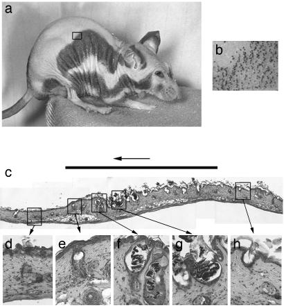
In the course of animal development, complex structures form autonomously from the apparently shapeless egg. How cells can produce spatial patterns that are much larger than each cell is one of the key issues in developmental biology. It has been suggested that spatial patterns in animals form through the same principles by which dispatched structures are formed in the nonbiological system. However, because of the complexity of biological systems, molecular details of such phenomena have been rarely clarified. In this article, we introduce an example of a pattern-forming phenomenon that occurs in the skin of mutant mice. The mutant mouse has a defect in splicing of the Foxn1 (Whn or nude) gene, which terminates hair follicle development just after pigment begins to accumulate in the follicle. The immature follicles are rapidly discharged, and a new hair cycle resumes. Eventually, the skin color of the mouse appears to oscillate. The color oscillation is synchronous in juvenile mice, but the phase gradually shifts among skin regions to eventually form traveling, evenly spaced stripes. Although the time scale is quite different, the pattern change in the mutant mouse shares characteristics with the nonlinear waves generated on excitable media, such as the Belousov-Zhabotinskii reaction, suggesting that a common principle underlies the wave pattern formation. Molecular details that underlie the phenomenon can be conjectured from recent molecular studies.


Figures

Fig. 1.
Fig. 1.
Stripe patterns of homozygous Foxn1tw mouse and skin sections. (a) Homozygous Foxn1tw mouse at 120 days after birth. (b) Close-up of the border of the pigmented region. The region shown is indicated by the square in a.(c) Paraffin section (×100) of skin cut from the side of the body and along the direction in which the wave runs. The thick line over the picture shows the region of a black stripe. The stripe is running leftward at a speed of ≈1.5 mm per 30 days. (dh) Paraffin section (×400) of each position of the wave. (Scale bars: 50 μm.)
Fig. 2.
Fig. 2.
Two types of aberrant alternative splicing between exons 6 and 7 of Foxn1tw mRNA. (Top) Normal splicing between exons 6 and 7 of Foxn1 mRNA. The nucleotide numbers of 5′ and 3′ of intron 6 are 39651 and 43956, respectively. The nucleotide numbers are referred to in the reported M. musculus whn gene sequence, GenBank accession no. Y12488. (Middle) The major form of the mutant mRNA. This type, 77% of the total of Foxn1tw mRNA, has a 54-bp deletion 5′ in exon 7 (43957–44010), generating an 18-aa deletion in the center of the DNA binding domain. (Bottom) The minor form of the mutant mRNA. This type, 23% of the total of Foxn1tw mRNA, has a 44-bp cryptic exon (40541–40584) and a 34-bp deletion 5′ in exon 7 (43957–43991), generating a translational termination (43992–43994).
Fig. 3.
Fig. 3.
Pattern change of a homozygous Foxn1tw mouse during the 30-day cycle. (a) Days 30–60. (b) Days 90–120. (c) Days 210–240 after birth. Pictures are taken at 5-day intervals with a Nikon digital camera. The pattern change shown here is typical for a homozygous Foxn1tw mouse.
Fig. 4.
Fig. 4.
Summary of stripe movement. (a and b) Side and top views of the adult Foxn1tw mouse, respectively. (c and d) Schematic drawing of typical wave movement in an adult Foxn1tw mouse. The numbers by each line represent the time course of a wave. The time interval between the numbers is ≈30 days. (e) Traveling waves formed by the BZ reaction. The shape of the mouse is superimposed. The outermost wave is the first wave. Intervals between waves are relatively wider for the first a few waves, then become shorter and constant. BZ waves are made by the standard method described in ref. . To indicate the centers of concentric waves, two iron grains were put on the dish.
Fig. 5.
Fig. 5.
Color patterns of cats, mice, and porcupines. (ac) Periodic color pattern of house cat, mouse, and African porcupine. (d) Outlook of African porcupine. (e) Synchronized hair pattern of quills of African porcupine. (f) Skin and the roots of quills. (g) Schematic drawing explaining the reason the hair pattern looks synchronized.

Comment in

  • How the mouse got its stripes.
    Maini PK. Maini PK. Proc Natl Acad Sci U S A. 2003 Aug 19;100(17):9656-7. doi: 10.1073/pnas.1734061100. Epub 2003 Aug 11. Proc Natl Acad Sci U S A. 2003. PMID: 12913120 Free PMC article. No abstract available.

References

    1. Ball, P. (1988) The Self-Made Tapestry (Oxford Univ. Press, New York).
    1. Glansdorff, P. & Prigogine, I. (1971) Thermodynamics of Structure, Stability, and Fluctuations (Wiley, London).
    1. Kareiva, P. (1984) in Springer Lecture Notes in Biomathematics, ed. Hallam, T. (Springer, Berlin), Vol. 54, pp. 368–389.
    1. Meinhardt, H. (1982) Models of Biological Pattern Formation (Academic, London).
    1. Murray, J. D. (1993) Mathematical Biology (Springer, Berlin).

Publication types

MeSH terms

LinkOut - more resources