. 2003 Aug 19;100(17):9873-7.
doi: 10.1073/pnas.1734000100. Epub 2003 Jul 31.

A SNARE required for retrograde transport to the endoplasmic reticulum

Affiliations

A SNARE required for retrograde transport to the endoplasmic reticulum

Lena Burri et al. Proc Natl Acad Sci U S A. .

Abstract

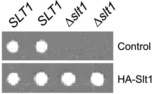
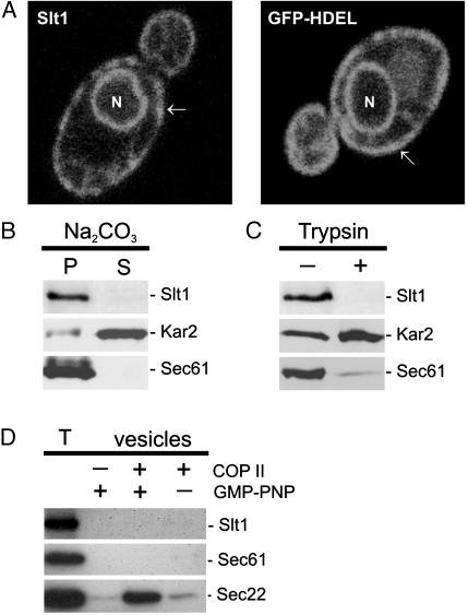
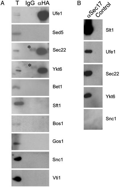
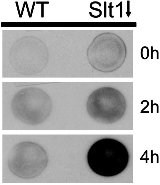
SNAREs (soluble N-ethylmaleimide-sensitive factor attachment protein receptors) are central components of the machinery mediating membrane fusion in all eukaryotic cells. Sequence analysis of the yeast genome revealed a previously uncharacterized SNARE, SNARE-like tail-anchored protein 1 (Slt1). Slt1 is an essential protein localized in the endoplasmic reticulum (ER). It forms a SNARE complex with Sec22 and the ER syntaxin Ufe1. Down-regulation of Slt1 levels leads to improper secretion of proteins normally resident in the ER. We suggest that Slt1 is a component of the SNAREpin required for retrograde traffic to the ER. Based on the previously reported association with Ufe1 and Sec22, Sec20 likely contributes the fourth SNARE to the SNAREpin.


Figures

Fig. 1.
Fig. 1.
(A) Amino acid sequence of Slt1. The SNARE motif is boxed in gray and the transmembrane domain is in boldface type. (B) Alignment of Slt1 and Sec20 with SNARE motif classes in yeast. Numbers denote residues in helix–helix contacts, the residue in the “0 layer” is boxed in black, and hydrophobic residues in the helix–helix contacts positions are shaded (–39).
Fig. 2.
Fig. 2.
Slt1 is an essential protein. SLT1slt1 yeast cells were transformed with a plasmid encoding HA-tagged Slt1 or a control plasmid. The cells were transferred to sporulation medium to induce meiosis, and the resulting spores were dissected onto plates of rich medium. Colonies were viewed after 3 days' growth at 30°C.
Fig. 3.
Fig. 3.
Slt1 is an integral membrane protein permanently localized in the ER. (A) Slt1 is localized to the ER. Yeast cells expressing GFP-Slt1 or GFP-HDEL (ER retention signal) were analyzed by confocal fluorescence microscopy. Both fusions target GFP to the ER (arrows, peripheral membrane; N, nucleus). (B) Slt1 is an integral membrane protein. A crude membrane fraction was isolated from yeast cells expressing GFP-Slt1, treated with 0.1M Na2CO3, and centrifuged to separate solubilized proteins (S) from insoluble material (P). (C) Slt1 is a type II membrane protein. The membrane fraction was incubated in the presence (+) or absence (–)of1.5 μg of trypsin. (D) Slt1 is not packaged into COPII vesicles. Isolated microsomes were incubated with purified Sar-1, Sec23/24, Sec13/31, and GMP-PNP as indicated. COP II vesicles were isolated as described in Material and Methods. T shows 10% of the microsomes in the budding reaction. Proteins were separated by SDS/PAGE and detected by Western blot analyses.
Fig. 4.
Fig. 4.
Slt1 forms a complex with SNAREs implicated in retrograde transport to the ER. (A) Detergent extracts of sec18-1 cells, which had been transformed with HA-Slt1, were incubated with an anti-HA affinity column, and coprecipitated proteins were detected by Western blot analysis. Asterisks indicate nonspecific IgG cross-reactivity. (B) Detergent extracts of sec18-1 cells, which had been transformed with HA-Slt1, were incubated with a GST-α-SNAP affinity column. Assembled v-/t-SNARE complexes were eluted with 1 M NaCl and immunoprecipitated with an anti-HA antibody, and Slt1-associated proteins were detected by Western blot analysis.
Fig. 5.
Fig. 5.
Inactivation of Slt1 results in the secretion of ER residents. Wild-type cells or Δslt1 cells expressing SLT1 under control of the GAL promoter were grown to log phase in rich medium with galactose as a carbon source (YPGal). To repress expression of SLT1, cells were spotted onto glucose plates (YPAD) and incubated at 30°C for 0, 2, or 4 h, then overlaid with nitrocellulose filters and allowed to grow at 30°C for an additional hour. Kar2 that had been secreted and bound to the nitrocellulose was detected by immunoblotting with an anti-Kar2 antibody.

References

    1. Söllner, T., Whiteheart, S. W., Brunner, M., Erdjument-Bromage, H., Geromanos, S., Tempst, P. & Rothman, J. E. (1993) Nature 362, 318–324. - PubMed
    1. Weber, T., Zemelman, B. V., McNew, J. A., Westermann, B., Gmachl, M., Parlati, F., Söllner, T. H. & Rothman, J. E. (1998) Cell 92, 759–772. - PubMed
    1. Sutton, R. B., Fasshauer, D., Jahn, R. & Brunger, A. T. (1998) Nature 395, 347–353. - PubMed
    1. Fukuda, R., McNew, J. A., Weber, T., Parlati, F., Engel, T., Nickel, W., Rothman, J. E. & Söllner, T. H. (2000) Nature 407, 198–202. - PubMed
    1. Melia, T. J., Weber, T., McNew, J. A., Fisher, L. E., Johnston, R. J., Parlati, F., Mahal, L. K., Söllner, T. H. & Rothman, J. E. (2002) J. Cell Biol. 158, 929–940. - PMC - PubMed

Publication types

MeSH terms