Review
. 2003 Aug 2;327(7409):280-3.
doi: 10.1136/bmj.327.7409.280.

Percutaneous interventional electrophysiology

Affiliations
Review

Percutaneous interventional electrophysiology

Gerry C Kaye. BMJ. .
No abstract available


Figures

Figure 1
Figure 1
Mechanism of a re-entry circuit. An excitation wave is propagated at a normal rate down path A, but slowly down path B. An excitation wave from an extrasystole now encounters the slow pathway (B), which is still refractory, creating unidirectional block. There is now retrograde conduction from path A, which coincides with the end of the refractory period in path B. This gives rise to a persistent circus movement
Figure 2
Figure 2
Classification of arrhythmias
Figure 3
Figure 3
Diagrams showing position of pacing or recording electrodes in the heart in the right anterior oblique and left anterior oblique views (views from the right and left sides of the chest respectively). HRA=high right atrial electrode, usually on the lateral wall or appendage; HBE=His bundle electrode, on the medial aspect of the tricuspid valve; RVA=right ventricular apex; CSE=coronary sinus electrode, which records electrical deflections from the left side of the heart between the atrium and ventricle
Figure 4
Figure 4
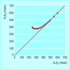
A normal atrioventricular nodal “hockey stick” curve during antegrade conduction of atrial extrastimuli. As the atrial extrastimulus (A1-A2) becomes more premature, the AH interval (H1-H2) shortens until the atrioventricular node becomes functionally refractory
Figure 5
Figure 5
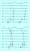
Coronary sinus electrode signals, with poles CS9-10 placed proximally near the origin of the coronary sinus and poles 1-2 placed distally reflecting changes in the left ventricular-left atrial free wall. Top: normal retrograde activation sequence with depolarisation passing from the ventricle back through the atrioventricular node to the right atrium and simultaneously across the coronary sinus to the left atrium. Bottom: retrograde activation sequence in the presence of an accessory pathway in the free wall of the left ventricle showing a shorter ventriculoatrial (VA) time than would be expected in the distal coronary sinus electrodes (CS1-2). Such a pathway would not be discernible from a surface electrocardiogram
Figure 6
Figure 6
Mechanisms for orthodromic (left) and antedromic (right) atrioventricular re-entrant tachycardia
Figure 7
Figure 7
Surface electrocardiogram leads V1 and V5 and signals from the distal coronary sinus electrodes (CS dist), proximal electrodes (CS prox), and the tip of the ablation catheter (ABL CATH) during pathway ablation to treat Wolff-Parkinson-White syndrome. The onset of radiofrequency energy (thin arrow) produces loss of pre-excitation after two beats with a narrow complex QRS seen at the fourth beat (broad arrow). Prolongation of the AV signal in the coronary sinus occurs when pre-excitation is lost
Figure 8
Figure 8
Atrioventricular nodal curves. In a patient with slow-fast junctional re-entrant tachycardia (left) there is a “jump” in atrioventricular nodal conduction when conduction changes from the fast to the slow pathway. In a patient with accessory pathways conducting antegradely (such as Wolff-Parkinson-White syndrome) there is no slowing of conduction as seen in the normal atrioventricular node, and the curve reflects conduction exclusively over the pathway (right)
Figure 9
Figure 9
Mechanism of slow-fast junctional re-entrant tachycardia. A premature atrial impulse finds the fast pathway refractory, allowing retrograde conduction back up to the atria
Figure 10
Figure 10
Diagram of basket-shaped mapping catheter with several recording electrodes (red dots). The basket retracts into a catheter for placement in either the atria or ventricles. Once it is in position, retraction of the catheter allows the basket to expand

References

    1. Olgin JE, Zipes DP. Specific arrhythmias: diagnosis and treatment. In: Braunwald E, Zipes DP, Libby P, eds. Heart disease. 6th ed. Philadelphia: Saunders, 2001: 1877-85
    1. McGuire MA, Janse MJ. New insights on the anatomical location of components of the reentrant circuit and ablation therapy for atrioventricular reentrant tachycardia. Curr Opin Cardiol 1995;10: 3-8 - PubMed
    1. Jackman WM, Beckman KJ, McClelland JH, Wang X, Friday KJ, Roman CA, et al. Treatment of supraventricular tachycardia due to atrioventricular nodal re-entry by radiofrequency catheter ablation of the slow-pathway conduction. N Engl J Med 1992;327: 313-8 - PubMed
    1. Calkins H, Leon AR, Deam AG, Kalbfleisch SJ, Langberg JJ, Morady F. Catheter ablation of atrial flutter using radiofrequency energy. Am J Cardiol 1994;73: 353-6 - PubMed
    1. Schilling RJ, Peter NS, Davies DW. Feasibility of a non-contact catheter for endocardial mapping of human ventricular tachycardia. Circulation 1999;99: 2543-52 - PubMed

MeSH terms