. 2003 Aug;185(16):4672-82.
doi: 10.1128/JB.185.16.4672-4682.2003.

Mutation in the relA gene of Vibrio cholerae affects in vitro and in vivo expression of virulence factors

Affiliations

Mutation in the relA gene of Vibrio cholerae affects in vitro and in vivo expression of virulence factors

Shruti Haralalka et al. J Bacteriol. 2003 Aug.

Abstract

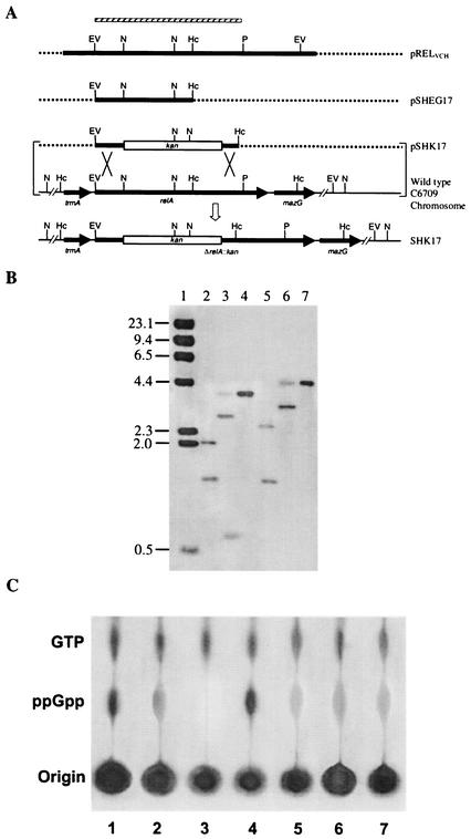
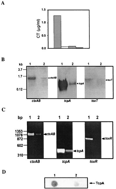
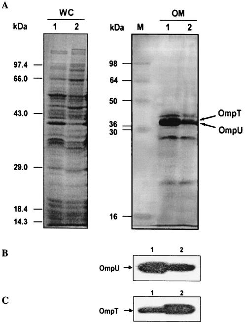
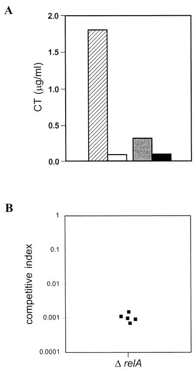
The relA gene product determines the level of (p)ppGpp, the effector nucleotides of the bacterial stringent response that are also involved in the regulation of other functions, like antibiotic production and quorum sensing. In order to explore the possible involvement of relA in the regulation of virulence of Vibrio cholerae, a relA homolog from the organism (relA(VCH)) was cloned and sequenced. The relA(VCH) gene encodes a 738-amino-acid protein having functions similar to those of other gram-negative bacteria, including Escherichia coli. A deltarelA::kan allele was generated by replacing approximately 31% of the open reading frame of wild-type relA of V. cholerae El Tor strain C6709 with a kanamycin resistance gene. The V. cholerae relA mutant strain thus generated, SHK17, failed to accumulate (p)ppGpp upon amino acid deprivation. Interestingly, compared to the wild type, C6709, the mutant strain SHK17 exhibited significantly reduced in vitro production of two principal virulence factors, cholera toxin (CT) and toxin-coregulated pilus (TCP), under virulence gene-inducing conditions. In vivo experiments carried out in rabbit ileal loop and suckling mouse models also confirmed our in vitro results. The data suggest that (p)ppGpp is essential for maximal expression of CT and TCP during in vitro growth, as well as during intestinal infection by virulent V. cholerae. Northern blot and reverse transcriptase PCR analyses indicated significant reduction in the transcript levels of both virulence factors in the relA mutant strain SHK17. Such marked alteration of virulence phenotypes in SHK17 appears most likely to be due to down regulation of transcript levels of toxR and toxT, the two most important virulence regulatory genes of V. cholerae. In SHK17, the altered expression of the two outer membrane porin proteins, OmpU and OmpT, indicated that the relA mutation most likely affects the ToxR-dependent virulence regulatory pathway, because it had been shown earlier that ToxR directly regulates their expression independently of ToxT.


Figures

FIG. 1.
FIG. 1.
Genetic organization of the relA region of the V. cholerae chromosome and comparison with those of different gram-negative organisms. The large arrows represent ORFs. The thick and thin lines represent intergenic regions and chromosomal DNA, respectively. The small arrows indicate the positions of the two primers, VCR-F and VCR-R, used to amplify the relA gene region (3.21 kb) of V. cholerae. The trmA gene codes for an RNA methyltransferase (23); mazG codes for a protein of unknown function in V. cholerae (23) and E. coli (38), but in S. enterica serovar Typhimurium it codes for a pyrophosphatase (38); STM2955, chpA, and chpR code for a PemK-like growth inhibitor, for a suppressor of ChpA, and for a putative transcriptional regulator, respectively.
FIG. 2.
FIG. 2.
(A) Schematic diagram (not to scale) showing the strategy for the construction of a relA deletion-insertion mutant strain, SHK17, of V. cholerae O1 El Tor. Restriction maps of the relA regions of the parental and mutant strains are shown. The thick lines represent gene regions, and the dashed lines represent vector DNA. The arrows indicate the direction of transcription of a gene. The thin lines indicate intergenic regions or chromosomal DNA. The hatched bar represents the EcoRV-PstI fragment of the relAVCH gene used as a probe in Southern hybridization studies, as shown in panel B. The plasmid pSHK17, containing the kanamycin resistance gene cassette (kan) within the relAVCH gene, was introduced into strain C6709 by conjugation, and the recombinant (shown by an open arrow), in which the ΔrelA::kan allele had replaced the wild-type relA, was isolated as described in Materials and Methods. Restriction enzyme sites: EV, EcoRV; Hc, HincII; N, NsiI; P, PstI. (B) Confirmation of the relA mutation in SHK17 as a double-crossover event by Southern analysis. The EcoRV-PstI fragment (shown in panel A) of the relA gene of V. cholerae was used for hybridization studies. Chromosomal DNA was digested with different restriction enzymes. For a detailed analysis of the autoradiogram, see the text. Lanes 2 to 4, wild-type C6709 DNA digested with HincII, NsiI, and EcoRV, respectively; lanes 5 to 7, SHK17 DNA digested with HincII, NsiI, and EcoRV, respectively. In lane 1, λ DNA digested with HindIII was run as a molecular size marker, and the sizes are indicated (in kilobase pairs) on the left. (C) Failure of (p)ppGpp accumulation in the relA mutant strain SHK17 upon amino acid starvation. 32Pi-labeled cells were grown either in MOPS-glucose minimal medium with amino acid starvation induced by the addition of 500 μg of SHMT/ml (lanes 1 to 5) or in rich medium (lanes 6 and 7), formic acid extracts of the cells were prepared, and aliquots were loaded on a polyethyleneimine-coated TLC plate. The spots were developed as described in Materials and Methods. Lanes: 1 to 3, E. coli strains CF1648 (wild type), CF1652 (ΔrelA), and CF1693 (ΔrelA ΔspoT), respectively; 4 and 6, V. cholerae strain C6709 (wild type); 5 and 7, SHK17 (ΔrelA::kan).
FIG. 3.
FIG. 3.
(A) CT production in V. cholerae C6709 and SHK17 under AKI conditions. CT was assayed by the GM1-ELISA method in the culture supernatant of C6709 (hatched bar) or SHK17 (open bar) or the sonicated cell lysate of C6709 (shaded bar) or SHK17 (solid bar). The data represent the average of three independent experiments, each done in duplicate. (B) Northern blot analysis of ctxAB, tcpA, and toxT, as indicated, in V. cholerae C6709 (lanes 1) and SHK17 (lanes 2). C6709 and SHK17 cells were grown under AKI conditions, and total cellular RNAs were prepared, electrophoresed, transferred to a nylon membrane, and hybridized with the ctxAB, tcpA, or toxT gene as shown. Molecular size markers are indicated on the left. (C) Detection of ctxAB, tcpA, and toxR transcripts of V. cholerae C6709 (lanes 1) and SHK17 (lanes 2) by RT-PCR. The strains were grown under AKI conditions, and total cellular RNAs were prepared and subjected to RT-PCR. The samples were run on a 1.5% agarose gel and visualized after being stained with ethidium bromide. DNA molecular size markers (HaeIII digests of φX174 DNA) are indicated on the left. (D) Immuno-dot-blot analysis to detect TcpA in V. cholerae C6709 (lane 1) and SHK17 (lane 2). Whole-cell lysates of the strains grown under AKI conditions were spotted on a polyvinylidene difluoride membrane and immunoblotted with anti-TcpA monoclonal antibody.
FIG. 4.
FIG. 4.
RelA affects the major outer membrane proteins in V. cholerae. (A) Whole-cell (WC) lysate or outer membrane (OM) fraction was prepared from the wild-type strain, C6709 (lanes 1), and its ΔrelA mutant, SHK17 (lanes 2). Equal amounts of protein samples of the strains were separated by SDS-PAGE (12.5% gel) and stained with Coomassie blue for visualization. M, molecular mass markers. (B and C) The OM fractions of C6709 (lanes 1) and SHK17 (lanes 2) were subjected to Western blot analysis using rabbit polyclonal antiserum against the V. cholerae protein OmpU (B) or OmpT (C).
FIG. 5.
FIG. 5.
In vivo CT production and intestinal colonization by V. cholerae C6709 and SHK17. (A) CT was measured in ileal loop fluid of C6709 (hatched bar) or SHK17 (open bar) or in sonicated cell lysate of C6709 (shaded bar) or SHK17 (solid bar). The data represent the average of five independent experiments, each done in duplicate. (B) RelA is required for intestinal colonization in V. cholerae. A suckling mouse assay was performed as described in Materials and Methods. The parental strain, C6709-R (C6709 Rifr), was coinoculated with SHK17 (ΔrelA Kmr). The competitive index is the ratio of output mutant to wild type (recovered from the small intestine) divided by the ratio of input mutant to wild type (inoculated into the mouse); thus, if a mutant strain has no colonization defect, the competitive index will be close to 1 (40). Each data point represents an individual mouse.

Similar articles

Cited by

References

    1. Altschul, S. F., T. L. Madden, A. A. Schaffer, J. Zhang, Z. Zhang, W. Miller, and D. J. Lipman. 1997. Gapped BLAST and PSI-BLAST: a new generation of protein database search programs. Nucleic Acids Res. 25:3389-3402. - PMC - PubMed
    1. Ausubel, F. M., R. Brent, R. E. Kingston, D. D. Moore, J. G. Seidman, J. A. Smith, and K. Struhl (ed.). 1989. Current protocols in molecular biology. John Wiley and Sons, New York, N.Y.
    1. Bhadra, R. K., S. Roychoudhury, R. K. Banerjee, S. Kar, R. Majumdar, S. Sengupta, S. Chatterjee, G. Khetawat, and J. Das. 1995. Cholera toxin (CTX) genetic element in Vibrio cholerae O139. Microbiology 141:1977-1983. - PubMed
    1. Bik, E. M., A. E. Bunschoten, R. J. Willems, A. C. Chang, and F. R. Mooi. 1996. Genetic organization and functional analysis of the otn DNA essential for cell-wall polysaccharide synthesis in Vibrio cholerae O139. Mol. Microbiol. 20:799-811. - PubMed
    1. Cashel, M. 1969. The control of ribonucleic acid synthesis in Escherichia coli. J. Biol. Chem. 244:3133-3141. - PubMed

Publication types

MeSH terms

Associated data

LinkOut - more resources