. 2003 Aug;185(16):4717-26.
doi: 10.1128/JB.185.16.4717-4726.2003.

Rod shape determination by the Bacillus subtilis class B penicillin-binding proteins encoded by pbpA and pbpH

Affiliations

Rod shape determination by the Bacillus subtilis class B penicillin-binding proteins encoded by pbpA and pbpH

Yuping Wei et al. J Bacteriol. 2003 Aug.

Abstract

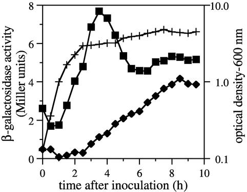
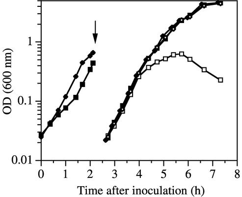
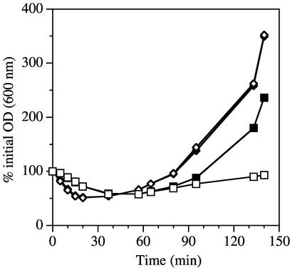
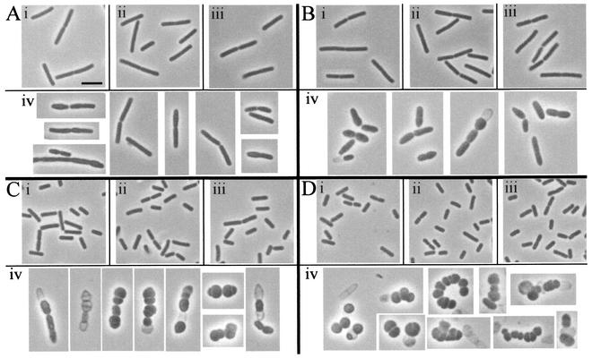
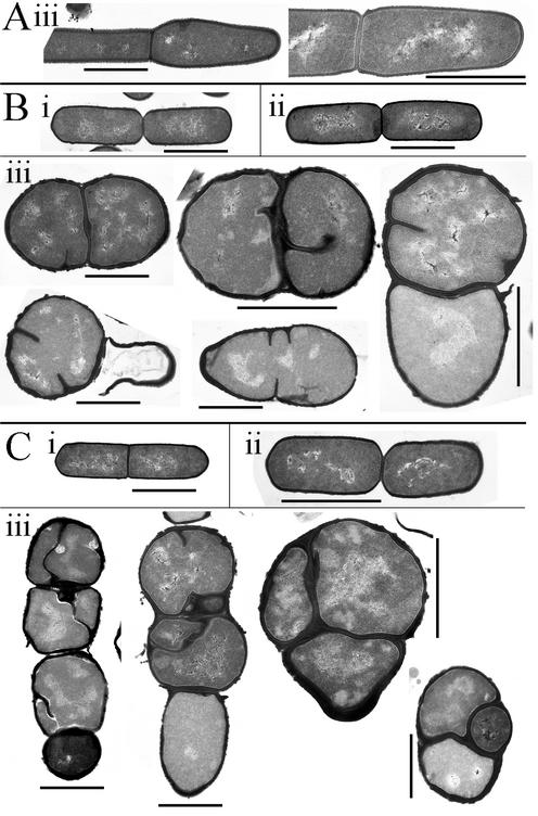
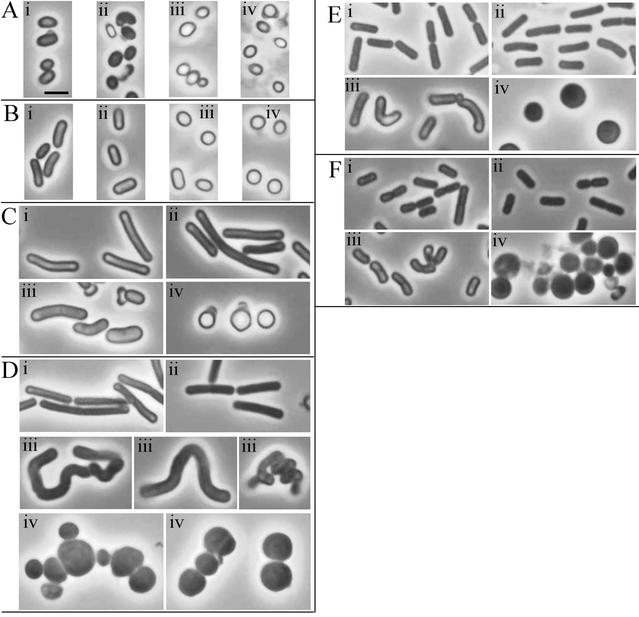
The peptidoglycan cell wall determines the shape and structural integrity of a bacterial cell. Class B penicillin-binding proteins (PBPs) carry a transpeptidase activity that cross-links peptidoglycan strands via their peptide side chains, and some of these proteins are directly involved in cell shape determination. No Bacillus subtilis PBP with a clear role in rod shape maintenance has been identified. However, previous studies showed that during outgrowth of pbpA mutant spores, the cells grew in an ovoid shape for several hours before they recovered and took on a normal rod shape. It was postulated that another PBP, expressed later during outgrowth, was able to compensate for the lack of the pbpA product, PBP2a, and to guide the formation of a rod shape. The B. subtilis pbpH (ykuA) gene product is predicted to be a class B PBP with greatest sequence similarity to PBP2a. We found that a pbpH-lacZ fusion was expressed at very low levels in early log phase and increased in late log phase. A pbpH null mutant was indistinguishable from the wild-type, but a pbpA pbpH double mutant was nonviable. When pbpH was placed under the control of an inducible promoter in a pbpA mutant, viability was dependent on pbpH expression. Growth of this strain in the absence of inducer resulted in conversion of the cells from rods to ovoid/round shapes and lysis. We conclude that PBP2a and PbpH play redundant roles in formation of a rod-shaped peptidoglycan cell wall.


Figures

FIG. 1.
FIG. 1.
Predicted pbpH transcription and translation start sites. (A) The N-terminal regions of five B. subtilis class B PBPs are aligned relative to their signal peptide hydrophobic cores (underlined) and several conserved residues immediately downstream of the signal peptides. The predicted PbpH N-terminal methionine residue (25) is indicated by an arrow. Several basic amino acids (shaded) would be incorporated upstream of the hydrophobic core if translation initiated 19 codons further upstream. (B) The DNA sequence begins with the start codon of a divergently oriented upstream gene, ykwD, and ends with the pbpH start codon predicted in the published genome sequence (25). The proposed alternative pbpH start codon is double underlined, and the putative ribosome-binding site is highlighted in gray. The apparent transcription start site at position 295 is indicated by an arrow, and potential σA recognition sequences are underlined. (C) RNA was purified from wild-type B. subtilis cells during early sporulation and used for reverse transcriptase primer extension (lane 1) with a 32P-, 5′-end-labeled primer that anneals within the pbpH sequence. Lane 2 is an identical primer extension reaction lacking RNA. DNA cycle sequencing reactions (lanes G, A, T, and C) utilized the same end-labeled primer. Due to the weak signal strength in lanes 1 and 2, the contrast of the image was adjusted independently for these lanes and for the sequencing lanes. The positions of the lanes relative to one another were not altered.
FIG. 2.
FIG. 2.
Expression of a pbpH-lacZ fusion. β-Galactosidase activity was determined in samples of cultures throughout growth and sporulation (sporulation initiated approximately 2.5 h after inoculation). Activity in the pbpH-lacZ strain DPVB168 (▪) was compared to the background activity found in the wild-type strain PS832 (⧫). The optical densities of the two cultures were essentially identical (+).
FIG. 3.
FIG. 3.
Growth is dependent on pbpH expression in the absence of pbpA. The wild-type strain PS832 (⧫ and ◊) and the pbpA pbpH amyE::xylAp-pbpH strain DPVB207 (▪ and □) were grown in the presence of xylose and then centrifuged (at the time indicated by the arrow), resuspended at a 20-fold dilution, and incubated in medium containing (solid symbols) or lacking (open symbols) xylose. OD, optical density.
FIG. 4.
FIG. 4.
Germination and outgrowth of spores expressing and lacking pbpH. Spores were produced from the wild-type strain PS832 (⧫ and ◊) and the pbpA pbpH amyE::xylAp-pbpH strain DPVB207 (▪ and □) in the presence of xylose and germinated in the presence (solid symbols) or absence (open symbols) of xylose.
FIG. 5.
FIG. 5.
Phase contrast microscopy of vegetative cells. Cultures were grown in the presence of xylose and then resuspended in medium containing or lacking xylose, as described for Fig. 3. Culture samples were examined 80 min (A), 120 min (B), 180 min (C), and 240 min (D) after resuspension. Cells are from the wild-type strain PS832 with (i) and without (ii) xylose and the pbpA pbpH amyE::xylAp-pbpH strain DPVB207 with (iii) and without (iv) xylose. All images are at the same magnification. Scale bar in Ai, 2 μm.
FIG. 6.
FIG. 6.
Electron microscopy of vegetative cells. Cultures were grown in the presence of xylose and then resuspended in medium containing or lacking xylose as described for Fig. 3. Culture samples were examined 80 min (A), 180 min (B), and 240 min (C) after resuspension. Cells are from the wild-type strain PS832 with xylose (i) and the pbpA pbpH amyE::xylAp-pbpH strain DPVB207 with (ii) and without (iii) xylose. Scale bars, 1 μm.
FIG. 7.
FIG. 7.
Phase contrast microscopy of germinating spores. Spores were germinated in medium containing or lacking xylose. Culture samples were examined 45 min (A), 60 min (B), 90 min (C), 120 min (D), 180 min (E), and 240 min (F) after resuspension. Cells are from the wild-type strain PS832 with (i) and without (ii) xylose and the pbpA pbpH amyE::xylAp-pbpH strain DPVB207 with (iii) and without (iv) xylose. All images are at the same magnification. Scale bar in Ai, 2 μm.

References

    1. Addinall, S. G., and J. Lutkenhaus. 1996. FtsZ-spirals and -arcs determine the shape of the invaginating septa in some mutants of Escherichia coli. Mol. Microbiol. 22:231-237. - PubMed
    1. Altschul, S. F., W. Gish, W. Miller, E. W. Myers, and D. J. Lipman. 1990. Basic local alignment search tool. J. Mol. Biol. 215:403-410. - PubMed
    1. Anagnostopoulos, C., and J. Spizizen. 1961. Requirements for transformation in Bacillus subtilis. J. Bacteriol. 81:74-76. - PMC - PubMed
    1. Anderson, A. J., R. S. Green, A. J. Sturman, and A. R. Archibald. 1978. Cell wall assembly in Bacillus subtilis: location of wall material incorporated during pulsed release of phosphate limitation, its accessibility to bacteriophages and concanavalin A, and its susceptibility to turnover. J. Bacteriol. 136:886-899. - PMC - PubMed
    1. Archibald, A. R., I. C. Hancock, and C. R. Harwood. 1993. Cell wall structure, synthesis, and turnover, p. 381-410. In A. L. Sonenshein, J. A. Hoch, and R. Losick (ed.), Bacillus subtilis and other gram-positive bacteria. American Society for Microbiology, Washington, D.C.

Publication types

MeSH terms

LinkOut - more resources