. 2003 Aug 1;17(15):1835-40.
doi: 10.1101/gad.266803.

Endothelial PDGF-B retention is required for proper investment of pericytes in the microvessel wall

Affiliations

Endothelial PDGF-B retention is required for proper investment of pericytes in the microvessel wall

Per Lindblom et al. Genes Dev. .

Abstract

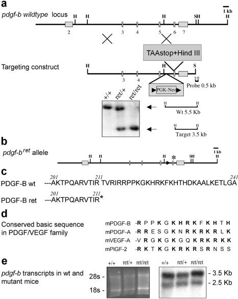
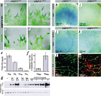
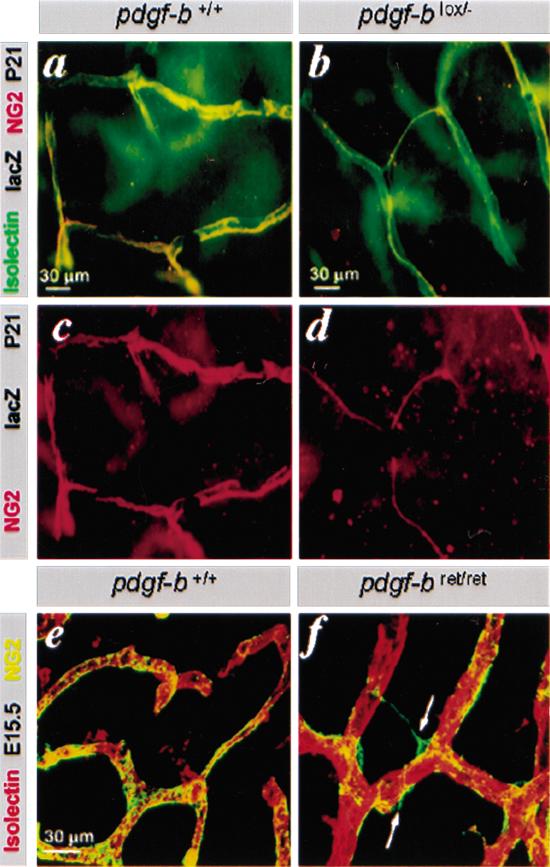
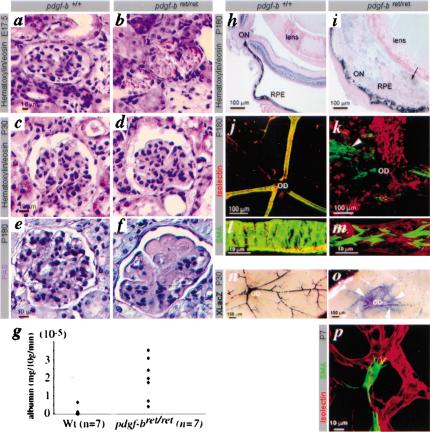
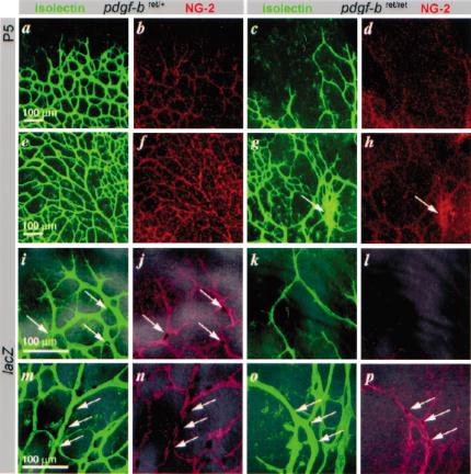
Several platelet-derived growth factor (PDGF) and vascular endothelial growth factor (VEGF) family members display C-terminal protein motifs that confer retention of the secreted factors within the pericellular space. To address the role of PDGF-B retention in vivo, we deleted the retention motif by gene targeting in mice. This resulted in defective investment of pericytes in the microvessel wall and delayed formation of the renal glomerulus mesangium. Long-term effects of lack of PDGF-B retention included severe retinal deterioration, glomerulosclerosis, and proteinuria. We conclude that retention of PDGF-B in microvessels is essential for proper recruitment and organization of pericytes and for renal and retinal function in adult mice.


Figures

Figure 1
Figure 1
The pdgf-bret allele. (a) Outline of the pdgf-b locus, targeting construct, and Southern identification of the pdgf-bret allele. (b) Schematic outline of the pdgf-bret allele. Remaining loxP site and inserted TAA-stop denoted by arrowhead and asterisk, respectively. (c) C-terminal sequence of the predicted PDGF-Bwt and PDGF-Bret proteins. (d) Comparison of the retention motifs of mouse PDGF and VEGF members. Basic residues in bold. (e) Northern blot analysis of pdgf-b transcripts in pdgf-b+/+, pdgf-bret/+, and pdgf-bret/ret brains. (Left) EtBr-stained gel.
Figure 2.
Figure 2.
Expression of the pdgf-bret allele, activity of the PDGF-Bret protein, and deficient recruitment of pericytes in pdgf-bret/ret mice. (a-d) In situ hybridization localizes pdgf-b mRNA to endothelial tip cells (arrows). (e,f) PDGF-B protein levels by proximity ligation assay in medium (M) and lysates (L) of wild-type (Wt) and pdgf-bret/ret (Ret) endothelioma cell lines. Triplicate measurements are shown with standard deviations. (g) PDGFR-β phosphorylation induced by the indicated dilutions (%) of 10× concentrated media from endothelioma cells. Western blots with phospho-tyrosine (P-tyr) and PDGFR-β (Rec-β) antibodies are shown. (h-k) XlacZ4 staining of forebrain from E15.5 pdgf-b+/+, pdgf-bret/+, pdgf-bret/ret, and pdgf-bret/- mice. LacZ-positive pericytes and vSMCs align cerebral/meningeal vessels. (l,m) GFAP (red) and isolectin (green) staining of P5 vibratome-sectioned brain. Up-regulation of GFAP in astrocytes is seen in focal regions of the pdgf-bret/ret brain.
Figure 3.
Figure 3.
Defective investment of pericytes into the vessel wall in pdgf-bret/ret mice. (a-d) Isolectin (green), NG2 (red), and LacZ stain of pdgf-b+/+ and pdgf-blox/- P21 retinas. The few pericytes seen in pdgfblox/- mice extend thin endothelium-associated processes. (e,f) NG2 and isolectin staining of E12.5 hindbrain. Pericytes (green) are partially detached and extend processes away from the endothelial cells (red; arrows) in pdgf-bret/ret mice.
Figure 4.
Figure 4.
Glomerulosclerosis, proteinuria, retinopathy, and vSMC deficiency in pdgf-bret/ret mice. H&E staining shows lack of mesangial cells in pdgf-bret/ret mice at E17.5 (a,b), normal histology at P30 (c,d), and glomerulosclerosis, by PAS staining at P180 (e,f). (g) Albuminuria in pdgf-bret/ret mice at 3 mo of age. (h-p) Analysis of postnatal retinas (P7-P180). (h-i) Retinal disorganization in pdgf-bret/ret mice, with fibrosis and invasion of RPE (arrow). (j,k) Isolectin (red) and SMA (green) staining of flat-mounted mouse retinas. Arrowhead indicates area of SMA-positive cells outside vessel walls. (l,m) Different density and organization of arterial SMCs in pdgf-b+/+ and pdgf-bret/ret retinas. (n,o) XlacZ4 staining of P30 retinas shows defective pericyte recruitment in pdgf-bret/ret mice and formation of vSMC sheets (arrowheads). (o) Arrows point at residual vessel-associated pericytes in peripheral regions. (p) Retina of P7 pdgf-bret/ret mouse with SMA-positive pericyte partially detached from the endothelium. ON, optic nerve; OD, optic disc.
Figure 5.
Figure 5.
Retinal vascular development in pdgf-bret/ret mice. Retinal vessels in control (pdgf-bret/+) and pdgf-bret/ret mice at P5. EC, isolectin (green); pericytes, NG2 (red); pericyte nuclei, XlacZ4 (dark blue, arrows). Peripheral (a,b) and central (e,f) regions in pdgf-bret/+ mice show formation of regular vascular plexuses. Note extensive coverage with pericytes (b,f), except for sprouting tips (b). In pdgfbret/ret mice, plexus spreading is delayed (c; peripheral), and irregular (g; central) with regions of hyperfusion (g, arrow), and reduced pericyte density (d,h). Peripheral (i-l) and central (m-p) regions at high magnification show that peripheral sprouting is sparse in pdgfbret/ret mice, leading to a wide-meshed, irregular vasculature partially devoid of pericytes. Few pericytes are present on remodeling arteries (cf. n and p, arrows).

References

    1. Allavena P., Dejana, E., Bussolino, F., Vecchi, A., and Mantovani, A. 1995. Cytokine regulation of endothelial cells. In Cytokines: A practical approach (ed. F.R. Balkwill), pp. 225-245. IRL Press, Oxford.
    1. Allt G. and Lawrenson, J.G. 2001. Pericytes: Cell biology and pathology. Cells Tissues Organs 169: 1-11. - PubMed
    1. Andersson M., Ostman, A., Westermark, B., and Heldin, C.H. 1994. Characterization of the retention motif in the C-terminal part of the long splice form of platelet-derived growth factor A-chain. J. Biol. Chem. 269: 926-9330. - PubMed
    1. Baeg G.H. and Perrimon, N. 2000. Functional binding of secreted molecules to heparin sulphate proteoglycans in Drosophila. Curr. Opin. Cell Biol. 12: 575-580. - PubMed
    1. Bussolino F., De Rossi, M., Sica, A., Colotta, F., Wang, J.M., Bocchietto, E., Padura, I.M., Bosia, A., Dejana, E., and Mantovani, A. 1991. Murine endothelioma cell lines transformed by polyoma middle T oncogene as target for and producers of cytokines. J. Immunol. 147: 2122-2129. - PubMed

Publication types

MeSH terms

Substances

LinkOut - more resources