. 2003 Aug 1;17(15):1909-20.
doi: 10.1101/gad.266203.

A conserved structural motif reveals the essential transcriptional repression function of Spen proteins and their role in developmental signaling

Affiliations

A conserved structural motif reveals the essential transcriptional repression function of Spen proteins and their role in developmental signaling

Mariko Ariyoshi et al. Genes Dev. .

Abstract

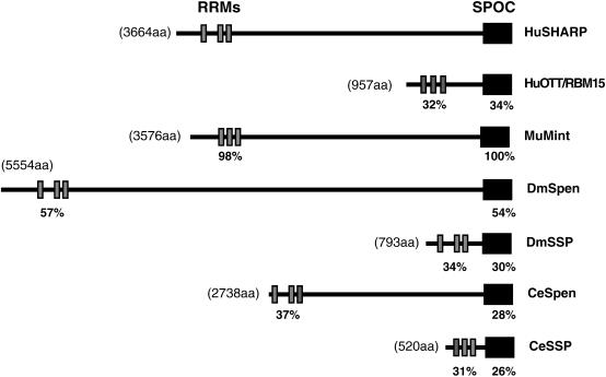
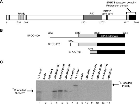
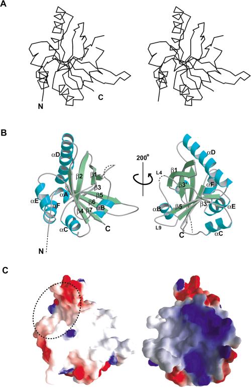
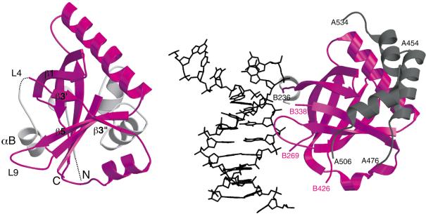
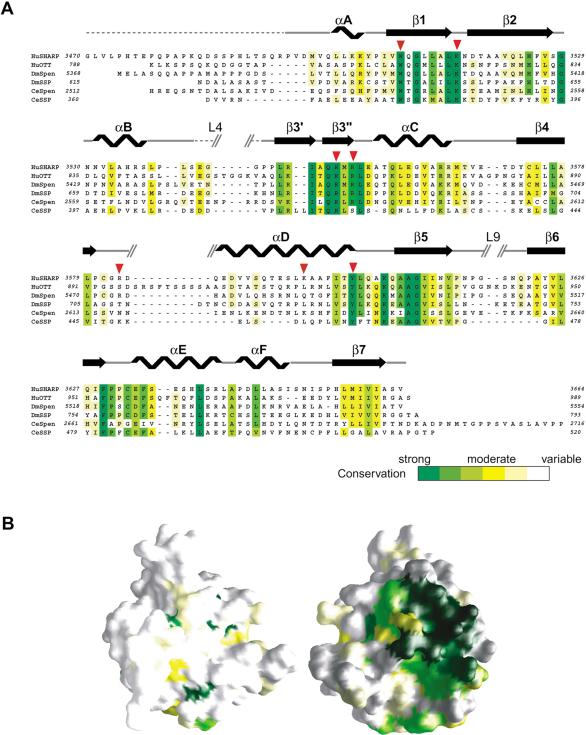
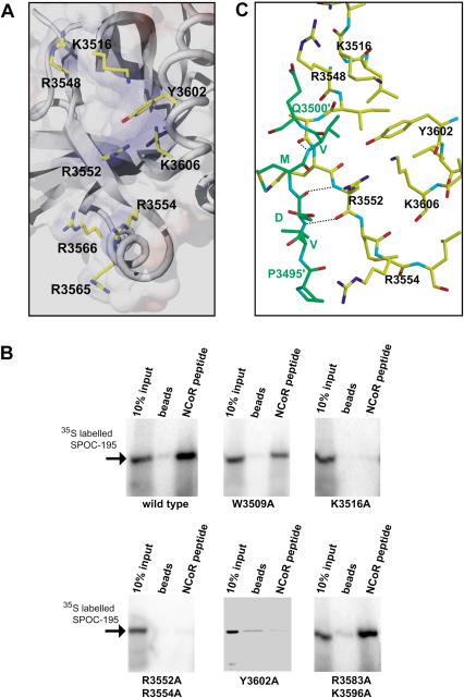
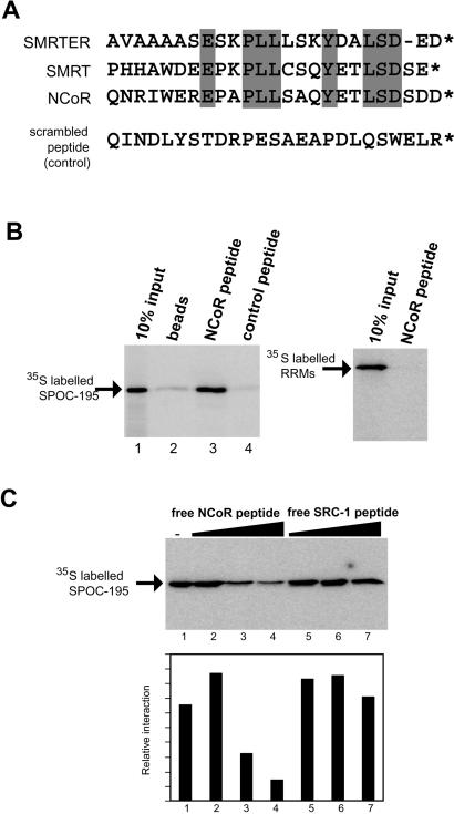
Spen proteins regulate the expression of key transcriptional effectors in diverse signaling pathways. They are large proteins characterized by N-terminal RNA-binding motifs and a highly conserved C-terminal SPOC domain. The specific biological role of the SPOC domain (Spen paralog and ortholog C-terminal domain), and hence, the common function of Spen proteins, has been unclear to date. The Spen protein, SHARP (SMRT/HDAC1-associated repressor protein), was identified as a component of transcriptional repression complexes in both nuclear receptor and Notch/RBP-Jkappa signaling pathways. We have determined the 1.8 A crystal structure of the SPOC domain from SHARP. This structure shows that essentially all of the conserved surface residues map to a positively charged patch. Structure-based mutational analysis indicates that this conserved region is responsible for the interaction between SHARP and the universal transcriptional corepressor SMRT/NCoR (silencing mediator for retinoid and thyroid receptors/nuclear receptor corepressor. We demonstrate that this interaction involves a highly conserved acidic motif at the C terminus of SMRT/NCoR. These findings suggest that the conserved function of the SPOC domain is to mediate interaction with SMRT/NCoR corepressors, and that Spen proteins play an essential role in the repression complex.


Figures

Figure 1.
Figure 1.
Common domain organization of the Spen proteins. HuSHARP, human SHARP; HuOTT, human OTT/RBM15; DmSpen, Drosophila Spen; DmSSP, Drosophila short Spen protein; CeSpen, C. elegans Spen; CeSSP, C. elegans short Spen protein. The approximate locations of RRMs and the SPOC domain of each protein are indicated in gray and black boxes, respectively. The percentages of identical residues in each domain of human OTT, Drosophila, and C. elegans homologs with human SHARP are indicated.
Figure 2.
Figure 2.
Functional domains of SHARP. (A) SHARP possesses a nuclear receptor interaction domain (RID) and RBP-Jκ interaction domain (RBPID) as well as RRMs and the SPOC domain. The C-terminal region of SHARP (amino acids 3417-3664), containing the SPOC domain, was characterized previously as a SMRT interaction domain or repression domain (Shi et al. 2001). (B) The recombinant C-terminal fragments of SHARP are shown schematically. The SPOC domain (amino acids 3498-3664) is represented by a black box. (C) The SPOC domain of SHARP interacts with C-SMRT. 35S-labeled in vitro translated C-SMRT (amino acids 2257-2517; lanes 1-7) and the N-terminal domain of PPARγ2 (amino acids 1-138; lanes 8-14) were incubated with purified GST or GST-SHARP fragments on glutathione beads. The bound proteins were analyzed by SDS-PAGE and visualized by fluorography. Note, there is a weak interaction between C-SMRT and the GST-RRMs. This is far weaker than the interaction with the GST-SPOC domain and seems unlikely to be biologically relevant.
Figure 3.
Figure 3.
Structure of the SPOC domain of SHARP. (A) Stereo view of Cα backbone structure of the SPOC domain. (B) Ribbon representation of structure of the SPOC domain (SPOC-195). Broken lines indicate the disordered regions at the N terminus and in the L4 loop (amino acids 3542-3544). The molecule on the right side is rotated by 200° around the vertical axis. (C) Electrostatic surface potential map of the SPOC domain. Red represents regions of negative charge, whereas blue shows positive charges. The molecules are in the same orientation as in B. A dotted circle indicates the hydrophobic and slightly acidic cavity described in text (left). The basic patch is located on the opposite side (right).
Figure 4.
Figure 4.
Structural similarity between the SPOC domain (left) and Ku80 β-barrel domain (PDB, 1jey; right). The structural elements in common are colored in magenta. The Ku80 domain is shown bound to DNA (black), and the three interacting helices from Ku70 are shown in gray. The long peptide loop (B269-B338) in Ku80, which wraps around DNA, is omitted for clarity.
Figure 5.
Figure 5.
Conservation of the basic cluster in Spen proteins. (A) Sequence conservation in Spen proteins. The sequence of SHARP SPOC domain, SPOC-195, was aligned with those of other spen proteins from human, Drosophila, and C. elegans using CLUSTALW (Thompson et al. 1994). Identical and similar residues are highlighted in a color scheme corresponding to conservation level as shown in the bar at bottom. The secondary structure elements of SPOC-195 are shown above the sequence. The strand β3 is divided into two parts: β3′ and β3′. Broken lines indicate the disordered regions. Red triangles indicate the mutation sites in the peptide interaction assay in Figure 7B. (B) Distribution of the conserved residues mapped on the surface of the SPOC domain. Residues are colored according to conservation as in A. The molecules are represented in the same orientation as in Figure 3. Note that the most conserved region, colored in green (right), corresponds to the basic cluster in Figure 3C.
Figure 7.
Figure 7.
The conserved basic cluster is essential for interaction with corepressor. (A) A view of the conserved basic region. Key residues are indicated by stick model beneath a semitransparent surface representation. (B) NCoR LSD peptide pull-down assay using wild-type and mutant SPOC domains. Mutation of the conserved tryptophan, W3509A, showed mildly reduced binding to corepressor. Mutations in conserved residues of the SPOC domain (K3516A, R3583A/K3596A, Y3620A) abolish interaction with corepressor. Mutations of nonconserved basic residues (R3583A/K3596A) had no effect. (C) Crystal packing interaction between the N terminus of one SPOC domain (Pro 3495-Gln 3500, in green) with the β3 strand of an adjacent molecule (Arg 3548-Arg 3554, in yellow). This may be indicative of the type of interaction with corepressor peptide. Backbone-backbone hydrogen bonds are indicated by broken lines.
Figure 6.
Figure 6.
Specific binding of the SPOC domain to the C-terminal peptide from the SMRT/NCoR corepressor. (A) The C-terminal sequence is well conserved in SMRT homologs. The sequences from Drosophila SMRT homolog (SMRTER), human SMRT, and human NCoR are compared. The conserved residues in the homologs are highlighted in gray. A scrambled peptide, which has the same amino acid composition as the NCoR peptide, but in a different order, was used as a control peptide. (B) Binding assay with corepressor peptide affinity beads. 35S-labeled SPOC-195 was incubated with NHS affinity beads linked to the C-terminal NCoR or control peptides. The SPOC domain is able to bind to the NCoR peptide (left, lane 3), but not with the control peptide. The RRMs from SHARP showed no binding (right). (C) Competition assay with free NCoR and coactivator SRC-1 peptides. The 35S-labeled SPOC domain bound to the NCoR peptide affinity beads was challenged with free NcoR peptide (lanes 2-4, using 10, 50, and 100 μM) and free SRC-1 peptide (lanes 5-7, using 5, 10, and 100 μM). Quantitation of the competition assay is shown at bottom.

References

    1. Bertrand N., Castro, D.S., and Guillemot, F. 2002. Proneural genes and the specification of neural cell types. Nat. Rev. Neurosci. 3: 517-530. - PubMed
    1. Brunger A.T., Adams, P.D., Clore, G.M., DeLano, W.L., Gros, P., Grosse-Kunstleve, R.W., Jiang, J.S., Kuszewski, J., Nilges, M., Pannu, N.S., et al. 1998. Crystallography & NMR system: A new software suite for macromolecular structure determination. Acta Crystallogr. D. Biol. Crystallogr. 54: 905-921. - PubMed
    1. Chen F. and Rebay, I. 2000. split ends, a new component of the Drosophila EGF receptor pathway, regulates development of midline glial cells. Curr. Biol. 10: 943-946. - PubMed
    1. Chen J.D. and Evans, R.M. 1995. A transcriptional co-repressor that interacts with nuclear hormone receptors. Nature 377: 454-457. - PubMed
    1. Collaborative Computational Project, Number 4. 1994. The CCP4 suite: Programs for protein crystallography. Acta Crystallogr. D. Biol. Crystallogr. 50: 760-763. - PubMed

Publication types

MeSH terms

LinkOut - more resources