. 2003 Aug 1;17(15):1921-32.
doi: 10.1101/gad.1099503.

BMAL1-dependent circadian oscillation of nuclear CLOCK: posttranslational events induced by dimerization of transcriptional activators of the mammalian clock system

Affiliations

BMAL1-dependent circadian oscillation of nuclear CLOCK: posttranslational events induced by dimerization of transcriptional activators of the mammalian clock system

Roman V Kondratov et al. Genes Dev. .

Abstract

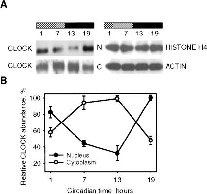
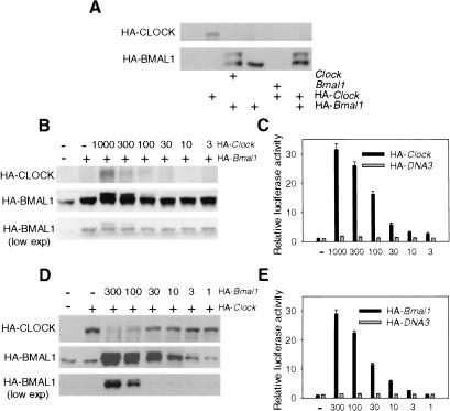
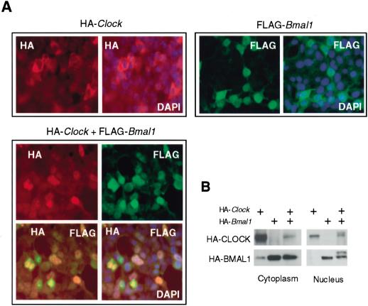
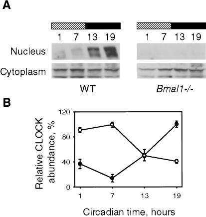
Mammalian CLOCK and BMAL1 are two members of bHLH-PAS-containing family of transcription factors that represent the positive elements of circadian autoregulatory feedback loop. In the form of a heterodimer, they drive transcription from E-box enhancer elements in the promoters of responsive genes. We have examined abundance, posttranslational modifications, cellular localization of endogenous and ectopically expressed CLOCK and BMAL1 proteins. Nuclear/cytoplasm distribution of CLOCK was found to be under circadian regulation. Analysis of subcellular localization of CLOCK in embryo fibroblasts of mice carrying different germ-line circadian mutations showed that circadian regulation of nuclear accumulation of CLOCK is BMAL1-dependent. Formation of CLOCK/BMAL1 complex following ectopic coexpression of both proteins is followed by their codependent phosphorylation, which is tightly coupled to CLOCK nuclear translocation and degradation. This binding-dependent coregulation is specific for CLOCK/BMAL1 interaction, as no other PAS domain protein that can form a complex with either CLOCK or BMAL1 was able to induce similar effects. Importantly, all posttranslational events described in our study are coupled with active transactivation complex formation, which argues for their significant functional role. Altogether, these results provide evidence for an additional level of circadian system control, which is based on regulation of transcriptional activity or/and availability of CLOCK/BMAL1 complex.


Figures

Figure 1.
Figure 1.
Circadian oscillation of CLOCK nuclear/cytoplasm distribution in the SCN. (A) SCN extracts obtained from C57BL/6J mice at designated circadian times were fractionated and analyzed by Western blotting using CLOCK antibodies. Histone H4 and β-actin were used for normalization. N, nuclear fraction; C, cytoplasm fraction. (B) Quantitative analysis of CLOCK protein expression in nuclear and cytoplasm fraction of the SCN. Data represent the average of three independent experiments. Each value represents a percentage of maximal level of CLOCK in nucleus or cytoplasm.
Figure 2.
Figure 2.
Effects of CLOCK and BMAL1 coexpression on their posttranslational modifications. (A) Western blot analysis of HEK-293 cells transfected with different combinations of Clock and Bmal1 expression plasmids. Dose dependence of HA-BMAL1 posttranslational modification (B), and transactivation of mPer1-Luc reporter (C) on HA-CLOCK amount. HEK-293 cells were transfected with constant amounts of pcHA-Bmal1 (100 ng), mPer1-Luc (50 ng), and pcDNA3-lacZ and indicated amount of pcHA-Clock. Dose dependence of HA-CLOCK abundance (D) and transactivation of mPer1-Luc reporter (E) on HA-BMAL1 amount. HEK-293 cells were transfected with constant amounts of pcHA-Clock (300 ng), mPer1-Luc (50 ng), and pcDNA3-lacZ and indicated amounts of pcHA-Bmal1. Expression of HA-CLOCK and HA-BMAL1 proteins in transfected cells were detected by Western blotting with anti-HA antibodies. Typical result from three independent experiments is presented. Note that in cells transfected by pcHA-Clock only, anti-HA antibodies at longer exposures recognize nonspecific band with electrophoretic mobility close to HA-BMAL1.
Figure 3.
Figure 3.
Nuclear/cytoplasm distribution of ectopically expressed CLOCK and BMAL1. (A) In situ detection of HA-CLOCK and Flag-BMAL1. HEK-293 cells were either individually transfected with pcHA-Clock and pcFlag-Bmal1 or cotransfected with both constructs. Secondary antibodies were conjugated with Texas Red (red fluorescence) for HA-CLOCK detection and FITC (green fluorescence) for Flag-BMAL1 detection. Orange color represents costaining of CLOCK and BMAL1 in cotransfected cells. DAPI (blue fluorescence) staining was used to visualize nuclei. (B) Western blot analysis of nuclear and cytoplasm fractions of HEK-293 cells transfected with pcHA-Clock and pcHA-Bmal1 in different combinations and probed with anti-HA antibodies.
Figure 4.
Figure 4.
Posttranslational modifications of CLOCK and BMAl1 are induced only by specific complex formation through the PAS domain. (A) Western blot analysis of nuclear and cytoplasm fractions of HEK-293 cells cotransfected by pcFlag-Bmal1 and HA-tagged CLOCK expression constructs (either full-length or deletion mutants). Anti-HA and anti-Flag antibodies were used for CLOCK and BMAL1 detection. Black asterisks designate bands corresponding to BMAL1 and full-length CLOCK; white asterisks designate bands corresponding to CLOCK truncated products of expected sizes. N, nucleus; C, cytoplasm. (B) Western blot analysis of HA-CLOCK and HA-BMAL1 ectopically expressed in HEK-293 cells in different combinations with Arnt, Ahr, and Hif1α expression plasmids and detected with anti-HA antibodies.
Figure 5.
Figure 5.
CLOCK nuclear localization is BMAL1-dependent. (A) Western blot analysis of CLOCK in primary fibroblasts derived from mouse embryos of different circadian genotypes. No CLOCK-specific immunoreactivity could be detected in Bmal1-/- MEFs. Histone H4 and β-actin were used for normalization of nuclear and cytoplasm fractions. (B) Western blot analysis of CLOCK temporal expression profile in wild-type and Bmal1-/- MEFs stimulated by serum shock. Serum was added at time point 0 and removed after 2 h. Two distinct peaks of CLOCK abundance in the nuclear fraction of wild-type fibroblasts are detected. An arrow indicates the position of CLOCK band. No CLOCK immunoreactivity is detected in the nuclear fraction of Bmal1-/- MEFs at all times tested (note that different exposure times used for detection of nuclear CLOCK in wild-type and Bmal1-/- fibroblasts). (C) Quantitative analysis of CLOCK nuclear/cytoplasm distribution in wild-type MEFs. •, nuclear CLOCK; ○, cytoplasmic CLOCK. Each value represents a percentage of the maximal level of CLOCK in the nucleus or the cytoplasm.
Figure 6.
Figure 6.
BMAL1 is required for circadian oscillation of nuclear CLOCK in mouse liver. (A) Western blot analysis of nuclear and cytoplasm CLOCK expression in livers of control wild-type and Bmal1-/- mice isolated at indicated circadian times. Note that longer exposure time was used for the nuclear fraction. (B) Quantitative analysis of CLOCK nuclear/cytoplasm distribution in livers of control mice. •, nuclear CLOCK; ○, cytoplasmic CLOCK. Mean data of three independent experiments are shown. Each value represents a percentage of the maximal level of CLOCK in the nucleus or the cytoplasm.
Figure 7.
Figure 7.
Schematic representation of posttranslational events induced by CLOCK/BMAL1 interaction. There are three possible scenarios: CLOCK/BMAL1 phosphorylation occurs in the cytoplasm and is required for BMAL1-dependent nuclear translocation of the complex (A); unphosphorylated CLOCK/BMAL1 are translocated to the nucleus (B,C), where they get phosphorylated either before (B) or after (C) initiation of transcription (see text for details).

References

    1. Balsalobre A., Damiola, F., and Schibler, U. 1998. A serum shock induces circadian gene expression in mammalian tissue culture cells. Cell 93: 929-937. - PubMed
    1. Balsalobre A., Marcacci, L., and Schibler, U. 2000. Multiple signaling pathways elicit circadian gene expression in cultured Rat-1 fibroblasts. Curr. Biol. 10: 1291-1294. - PubMed
    1. Camacho F., Cilio, M., Guo, Y., Virshup, D.M., Patel, K., Khorkova, O., Styren, S., Morse, B., Yao, Z., and Keesler, G.A. 2001. Human casein kinase Iδ phosphorylation of human circadian clock proteins period 1 and 2. FEBS Lett. 489: 159-165. - PubMed
    1. Dikstein R., Ruppert, S., and Tjian, R. 1996. TAFII250 is a bipartite protein kinase that phosphorylates the base transcription factor RAP74. Cell 84: 781-790. - PubMed
    1. Dolwick K.M., Swanson, H.I., and Bradfield, C.A. 1993. In vitro analysis of Ah receptor domains involved in ligand-activated DNA recognition. Proc. Natl. Acad. Sci. 90: 8566-8570. - PMC - PubMed

Publication types

MeSH terms