. 2003 Aug;23(16):5502-15.
doi: 10.1128/MCB.23.16.5502-5515.2003.

Degradation of normal mRNA in the nucleus of Saccharomyces cerevisiae

Affiliations

Degradation of normal mRNA in the nucleus of Saccharomyces cerevisiae

Biswadip Das et al. Mol Cell Biol. 2003 Aug.

Abstract

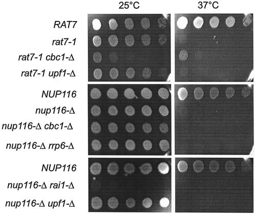
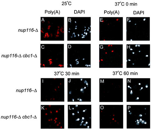
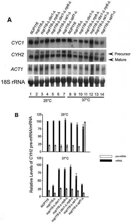
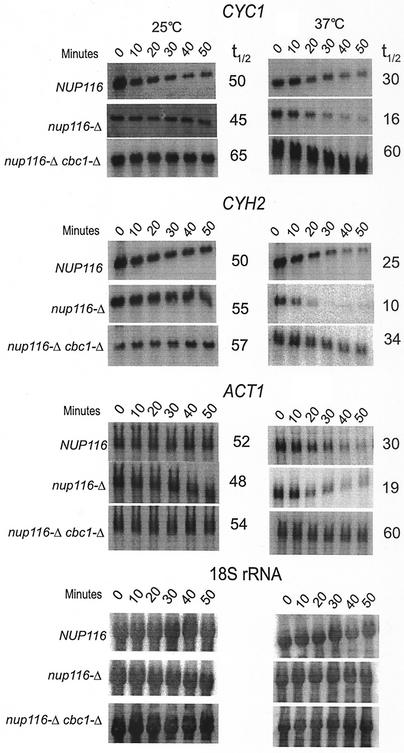
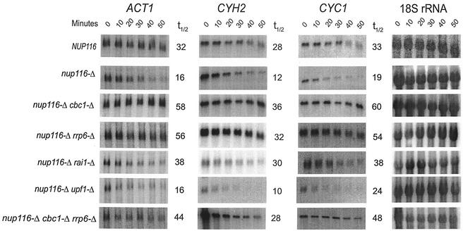
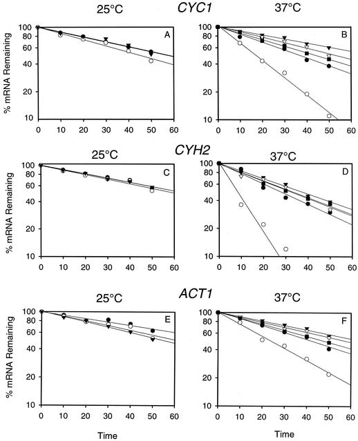
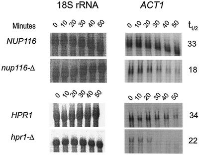
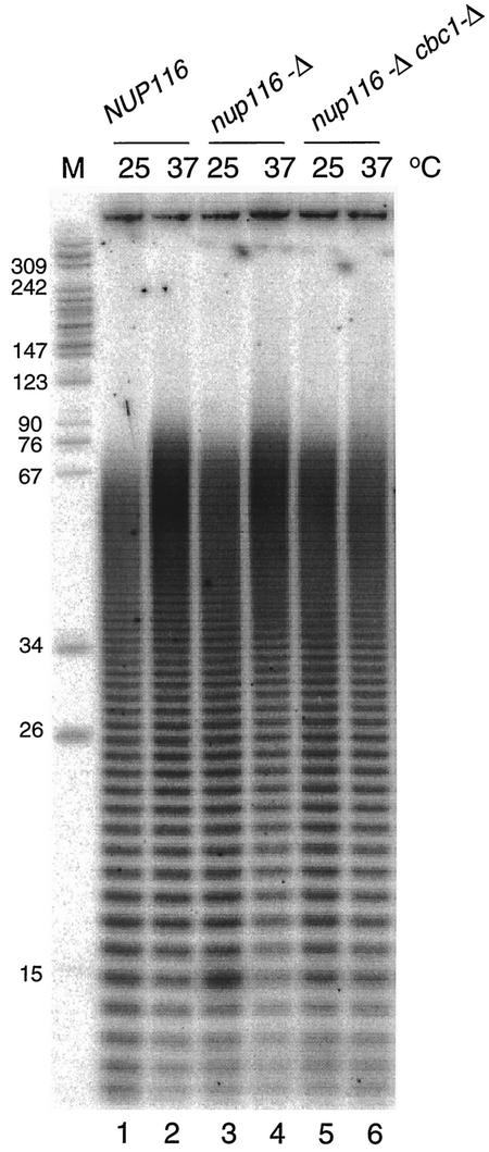
A nuclear mRNA degradation (DRN) system was identified from analysis of mRNA turnover rates in nup116-Delta strains of Saccharomyces cerevisiae lacking the ability to export all RNAs, including poly(A) mRNAs, at the restrictive temperature. Northern blotting, in situ hybridization, and blocking transcription with thiolutin in nup116-delta strains revealed a rapid degradation of mRNAs in the nucleus that was suppressed by the rrp6-delta, rai1-delta, and cbc1-delta deletions, but not by the upf1-delta deletion, suggesting that DRN requires Rrp6p, a 3'-to-5' nuclear exonuclease, the Rat1p, a 5'-to-3' nuclear exonuclease, and Cbc1p, a component of CBC, the nuclear cap binding complex, which may direct the mRNAs to the site of degradation. We propose that certain normal mRNAs retained in the nucleus are degraded by the DRN system, similar to degradation of transcripts with 3' end formation defects in certain mutants.


Figures

FIG. 1.
FIG. 1.
The growth of 1/10 serial dilutions of suspensions of various strains (Table 1) at 25°C for 4 days or 37°C for 2 days on YPD medium, demonstrating that the growth defect at 37°C of nup116-Δ strains is not suppressed by cbc1-Δ, rrp6-Δ, or other mutations, whereas the growth defect at 37°C of rat7-1 strains are partially suppressed by cbc1-Δ.
FIG. 2.
FIG. 2.
FISH analyses revealing that total poly(A)+ RNA in nup116-Δ and nup116cbc1-Δ strains are retained in the nucleus and that there is less degradation in the nup116cbc1-Δ strain. The isogenic pairs of strains were grown at 25°C to the mid-logarithmic phase of growth. Subsequently, one-half of each culture was transferred to the restrictive temperature of 37°C; the cultures were further incubated for one additional hour at both temperatures. Transcription of the cells in the culture shifted to 37°C was inhibited by the addition of thiolutin (4 μg/ml); the cells were harvested at various times after transcription block as indicated on top of each panel in the figure and subsequently fixed and processed for FISH and DAPI analysis as described in Materials and Methods. Left panels of mock shifted as well as each time point after transcription block show the localization and decay of the total poly(A)+ RNA as visualized using Cy3-labeled oligo(dT) are denoted as Poly(A), whereas right panels of respective time points show the nuclear DNA as visualized using DAPI staining denoted as DAPI. The time indicated at the top of each panel represents the time after transcription block to after shift to 37°C. See the Results (“Existence of DRN, a Cbc1p-dependent nuclear mRNA degradation pathway: cytological evidence”) for details of each panel.
FIG. 3.
FIG. 3.
Comparison of the steady-state levels of CYC1, ACT1, and CYH2 precursor and matured mRNAs at 25°C (mock-shifted) and 37°C (shifted) in NUP116 (normal), nup116-Δ, nup116cbc1-Δ, nup116rrp6-Δ, nup116cbc1rrp6-Δ, nup116rai1-Δ, and nup116upf1-Δ deletion strains. (A) Northern blots of steady-state levels of CYC1, ACT1, and CYH2 mRNAs and pre-mRNA in different strains are indicated at the top of each lane. All the strains were grown at 25°C until mid-log phase, half of the culture of each strain was then shifted to 37°C. Both cultures of each strain at 25 and 37°C were incubated for 1 h at the respective temperature and harvested. Subsequently, the steady-state levels of the each mRNA and pre-mRNA were determined by Northern blot analysis with the total RNA isolated from each of these strains and probing for respective pre-mRNA and mRNAs isolated from strains grown at 25°C (lanes 1 to 7) and shifted to 37°C (lanes 8 to 14). The signal for each mRNA in each lane was quantified as described in Materials and Methods and normalized against the 18S rRNA signals (shown at the lowest panel of A) for loading errors and the relative steady-state levels of each mRNA are presented in Table 2. (B) Quantification of the CYH2 pre-mRNA and mRNA signals. The intensity of each band of mature and precursor mRNA were determined by scanning the blots with a PhosphorImager and by normalizing for loading differences with respect to 18S rRNA signals. The relative levels of pre-mRNA and mRNA in each strain were expressed with respect to that in the NUP116 (normal) strain at each temperature, which was considered to be 100%. The error bar represents the range of three independent experiments.
FIG. 4.
FIG. 4.
Northern blot analysis revealing an increased degradation of CYC1, CYH2, and ACT1 mRNAs that are retained in the nucleus because of the export deficiency caused by nup116-Δ mutation. Furthermore, the Northern blot analysis also revealed that the degradation is suppressed by cbc1-Δ at 37°C. The NUP116 (normal), nup116-Δ, and nup116cbc1-Δ strains were grown at 25°C to the mid-logarithmic phase of growth. Subsequently, one-half of each culture was transferred to the restrictive temperature of 37°C. Both the cultures of each strain at 25 and 37°C were further incubated for one additional hour at both temperatures, and transcription was inhibited by the addition of thiolutin (4 μg/ml), as described in Materials and Methods. Cells of each strain from both the temperatures, mock shifted and shifted, were harvested after various times of thiolutin addition; Northern blots were prepared with total RNA; the half-lives of CYC1, CYH2, and ACT1 mRNA were determined as described in Materials and Methods and normalized against 18S rRNA shown at the bottom of the figure. The half-lives are presented beside each panel as well as in Table 3.
FIG. 5.
FIG. 5.
A comparison of the decay of ACT1, CYH2, and CYC1 mRNAs in NUP116 and various nup116-Δ strains by Northern blot analysis. The analysis was performed as described in the legend of Fig. 4 after normalizing each signal against that from 18S rRNA internal control (as shown on the rightmost panels) for each strain and the half-lives are presented beside each panel as well as in Table 4.
FIG. 6.
FIG. 6.
Graphical representation of decay of CYC1, CYH2 and ACT1 mRNAs at 25°C (A, C, and E) and at 37°C (B, D, and F) from thiolutin treated cells of NUP116 (•), nup116-Δ (○), nup116-Δ cbc1-Δ (▾), nup116-Δ rrp6-Δ (▿), and nup116-Δ cbc1-Δ rrp6-Δ (▪). The decay was determined by Northern blot analysis of the RNA extracted from the strains mentioned above treated with thiolutin from 0 to 50 min. The result from one typical experiment from each strain at different temperatures are presented as the percentage of mRNA remaining versus time of incubation of thiolutin.
FIG. 7.
FIG. 7.
Northern blot analysis revealing an increased degradation of ACT1 mRNA (right panels), which is retained in the nucleus because of the export deficiency caused by nup116-Δ or hpr1-Δ mutations at the restrictive temperature of 37°C when compared to the corresponding isogenic normal strain. The NUP116 (normal), nup116-Δ, and HPR1 and hpr1-Δ strains were grown at 25°C to the mid-logarithmic phase of growth. Subsequently, one-half of each culture was transferred to the restrictive temperature of 37°C; the cultures were further incubated for one additional hour at that temperature; and transcription was inhibited by the addition of thiolutin (4 μg/ml), as described in Materials and Methods. Northern blots were prepared using total RNA extracted from cells after various times, 0 to 50 min, of thiolutin addition. The half-lives, presented in Table 4, were determined from these blots after normalization to the 18S rRNA signals shown at the left panels. The numbers beside each panel represents the half-lives in minutes.
FIG. 8.
FIG. 8.
Poly(A) tail analysis of mRNA from strains carrying the nup116-Δ mutation. Poly(A) tails were analyzed by 3′ end labeling of 1 μg of total RNA with [32P]CP and RNA ligase, followed by hydrolysis with RNase A and RNase T1, electrophoretic separation on a 16% acrylamide-8 M urea gel, and storage phosphorimager analysis. Lane M indicates pBR322 MspI-cut length markers.

References

    1. Allmang, C., J. Kufel, G. Chanfreau, P. Mitchell, E. Petfalski, and D. Tollervey. 1999. Functions of the exosome in rRNA, snoRNA and snRNA synthesis. EMBO J. 18:5399-5410. - PMC - PubMed
    1. Allmang, C., E. Petfalski, A. Podtelejnikov, M. Mann, D. Tollervey, and P. Mitchell. 1999. The yeast exosome and human PM-Scl are related complexes of 3′→5′ exonucleases. Genes Dev. 13:2148-2158. - PMC - PubMed
    1. Bailer, S. M., C. Balduf, J. Katahira, A. Podtelejnikov, C. Rollenhagen, M. Mann, N. Pante, and E. Hurt. 2000. Nup116p associates with the Nup82p-Nsp1p-Nup159p nucleoporin complex. J. Biol. Chem. 275:23540-23548. - PubMed
    1. Beelman, C. A., A. Stevens, G. Caponigro, T. E. LaGrandeur, L. Hatfield, D. M. Fortner, and R. Parker. 1996. An essential component of the decapping enzyme required for normal rates of mRNA turnover. Nature 382:642-646. - PubMed
    1. Boeck, R., B. Lapeyre, C. E. Brown, and A. B. Sachs. 1998. Capped mRNA degradation intermediates accumulate in the yeast spb8-2 mutant. Mol. Cell. Biol. 18:5062-5072. - PMC - PubMed

Publication types

MeSH terms