. 2003 Aug;23(16):5516-25.
doi: 10.1128/MCB.23.16.5516-5525.2003.

Identification of a unique core domain of par-4 sufficient for selective apoptosis induction in cancer cells

Affiliations

Identification of a unique core domain of par-4 sufficient for selective apoptosis induction in cancer cells

Nadia El-Guendy et al. Mol Cell Biol. 2003 Aug.

Abstract

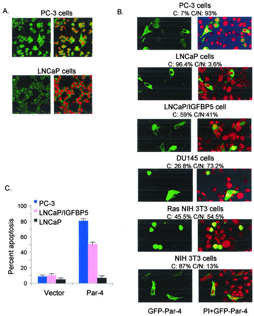
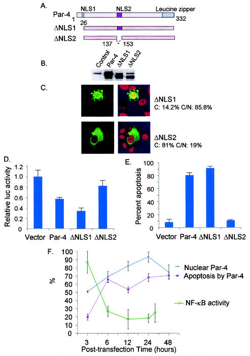
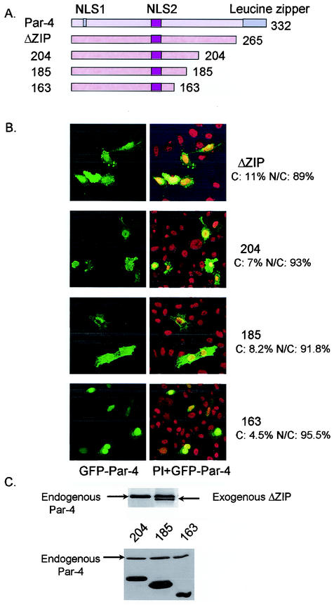
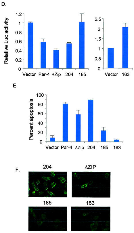
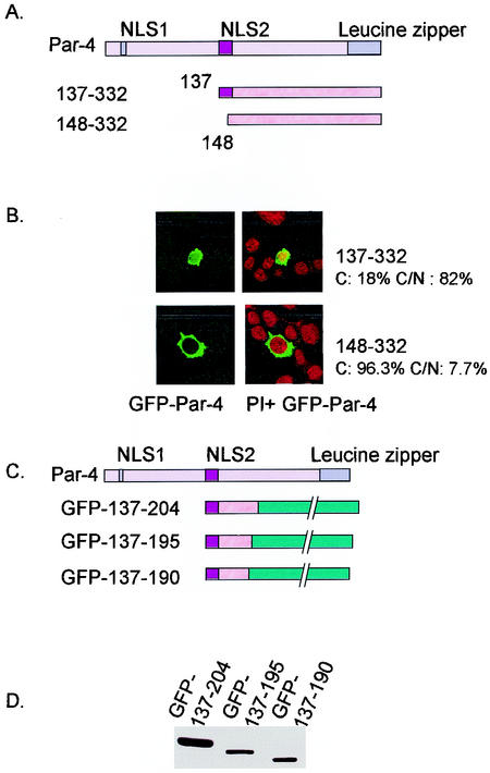
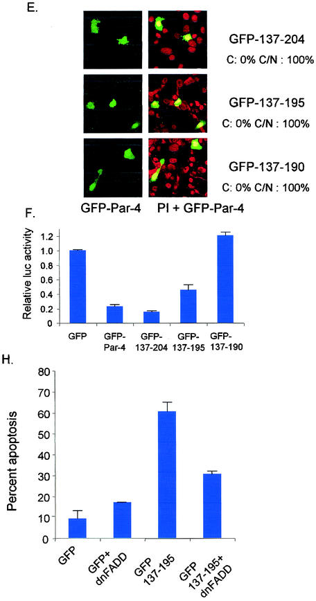
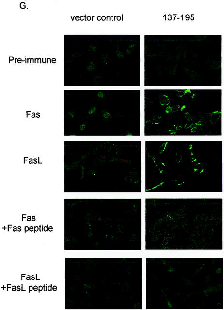
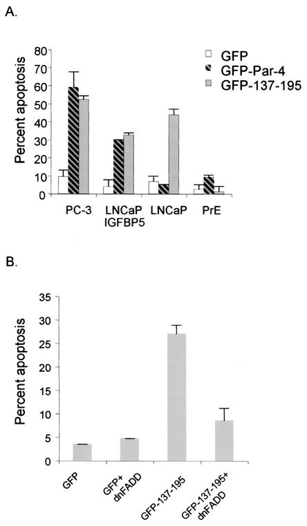
Recent studies indicated that the leucine zipper domain protein Par-4 induces apoptosis in certain cancer cells by activation of the Fas prodeath pathway and coparallel inhibition of NF-kappaB transcriptional activity. However, the intracellular localization or functional domains of Par-4 involved in apoptosis remained unknown. In the present study, structure-function analysis indicated that inhibition of NF-kappaB activity and apoptosis is dependent on Par-4 translocation to the nucleus via a bipartite nuclear localization sequence, NLS2. Cancer cells that were resistant to Par-4-induced apoptosis retained Par-4 in the cytoplasm. Interestingly, a 59-amino-acid core that included NLS2 but not the C-terminal leucine zipper domain was necessary and sufficient to induce Fas pathway activation, inhibition of NF-kappaB activity, and apoptosis. Most important, this core domain had an expanded target range for induction of apoptosis, extending to previously resistant cancer cells but not to normal cells. These findings have identified a unique death-inducing domain selective for apoptosis induction in cancer cells (SAC domain) which holds promise for identifying key differences between cancer and normal cells and for molecular therapy of cancer.


Figures

FIG. 1.
FIG. 1.
Nuclear localization of Par-4 correlates with apoptosis induction. PC-3 and LNCaP cells were fixed and probed with Par-4 antibody to detect expression of endogenous Par-4. Nuclei were visualized by staining with propidium iodide. Intracellular localization of endogenous Par-4 was recorded by confocal microscopy. Par-4 images are shown in the left panels, and overlay of Par-4 and propidium iodide images are shown in the right panels (A). Cells were transfected with GFP-Par-4 and treated with propidium iodide to detect nuclei. Intracellular localization of GFP-Par-4 was recorded by confocal microscopy (B). GFP-Par-4 images are shown in the left panels, and overlay of GFP-Par-4 and propidium iodide images are shown in the right panels. Note yellow color resulting from colocalization of GFP green fluorescence with reddish-orange propidium iodide staining (right panels), indicating nuclear expression of Par-4. The percentage of cells with strictly cytoplasmic and with cytoplasmic and nuclear Par-4 is indicated above each panel as C and C/N, respectively. To determine percent apoptosis, cells were transfected with GFP-Par-4 and the GFP vector as a control and subjected to DAPI staining. Apoptotic cells were quantified and expressed as a percentage of the total number of transfected cells (C).
FIG. 2.
FIG. 2.
NLS2 is required for nuclear entry, inhibition of NF-κB activity, and induction of apoptosis. Schematic representation of full-length Par-4, ΔNLS1, and ΔNLS2 is shown in panel A. PC-3 cells were transiently transfected with the vector control, Par-4, ΔNLS1, and ΔNLS2 and their GFP-tagged derivatives. Expression of endogenous Par-4 (control), ectopic Par-4, ΔNLS1, and ΔNLS2 proteins was examined by Western blot analysis (B). Intracellular localization of ΔNLS1 and ΔNLS2 (C) and their ability to induce apoptosis (E) were examined as indicated in the legend to Fig. 1. The percentage of cytoplasmic and cytoplasmic and nuclear Par-4 and mutant protein is indicated in panel C. Also note the apoptotic morphology of the ΔNLS1-transfected cell in the same panel. To determine inhibition of NF-κB transcriptional activity by Par-4 and its mutants (D), cells were transfected for 48 h with the RelA reporter system and β-galactosidase expression plasmid together with vector, Par-4, and mutant constructs. Whole-cell lysates were subjected to luciferase assays, and luciferase activity was normalized to the corresponding β-galactosidase activity. Luciferase activity by Par-4 and its mutants is expressed relative to the activity noted with the vector. The relative time course of nuclear translocation of ectopic Par-4, inhibition of NF-κB transcriptional activity, and induction of apoptosis is shown in panel F.
FIG. 3.
FIG. 3.
Leucine zipper domain is not essential for apoptosis. PC-3 cells were transiently transfected with untagged and GFP-tagged derivatives of various C-terminal deletion mutants of Par-4 (A). After 48 h, the cells were examined for intracellular localization of the mutants (B), expression of the mutant proteins by Western blot analysis (C), inhibition of NF-κB transcription activity (D), and apoptosis (E), as described in the legend to Fig. 1. The percentage of cells with strictly cytoplasmic and cytoplasmic and nuclear Par-4 mutant protein is indicated in panel B. To examine Fas translocation (F), PC-3 cells were transiently transfected with untagged C-terminal deletion mutants; after 48 h they were stained with anti-Fas antibody, and Alexa Fluor 488 green fluorescence was visualized by confocal microscopy.
FIG. 3.
FIG. 3.
Leucine zipper domain is not essential for apoptosis. PC-3 cells were transiently transfected with untagged and GFP-tagged derivatives of various C-terminal deletion mutants of Par-4 (A). After 48 h, the cells were examined for intracellular localization of the mutants (B), expression of the mutant proteins by Western blot analysis (C), inhibition of NF-κB transcription activity (D), and apoptosis (E), as described in the legend to Fig. 1. The percentage of cells with strictly cytoplasmic and cytoplasmic and nuclear Par-4 mutant protein is indicated in panel B. To examine Fas translocation (F), PC-3 cells were transiently transfected with untagged C-terminal deletion mutants; after 48 h they were stained with anti-Fas antibody, and Alexa Fluor 488 green fluorescence was visualized by confocal microscopy.
FIG. 4.
FIG. 4.
Identification of core domain of Par-4 that is sufficient for apoptosis. PC-3 cells were transiently transfected with GFP-tagged derivatives of Par-4 deletion mutants 137-332 and 148-332 (A) and examined for intracellular localization (B). Cells were transiently transfected with various deletion mutants of Par-4, 137-204, 137-195, and 137-190, and their GFP-tagged derivatives (C) and examined for expression by Western blot analysis (D), intracellular localization (E), inhibition of NF-κB activity (F), and apoptosis induction in the presence and absence of ectopic dominant-negative FADD (dnFADD) (H), as described in the legend to Fig. 1. The percentage of cells with strictly cytoplasmic and cytoplasmic and nuclear Par-4 mutant protein is indicated in panels B and E. To study Fas and FasL trafficking to the cell membrane, PC-3 cells were transfected with the 137-195 expression construct and the control vector for 48 h. The cells were then subjected to immunofluorescent staining with Fas and FasL antibody, preimmune normal rabbit antibody, Fas antibody preabsorbed with Fas peptide, and FasL antibody preabsorbed with FasL peptide as controls (G).
FIG. 4.
FIG. 4.
Identification of core domain of Par-4 that is sufficient for apoptosis. PC-3 cells were transiently transfected with GFP-tagged derivatives of Par-4 deletion mutants 137-332 and 148-332 (A) and examined for intracellular localization (B). Cells were transiently transfected with various deletion mutants of Par-4, 137-204, 137-195, and 137-190, and their GFP-tagged derivatives (C) and examined for expression by Western blot analysis (D), intracellular localization (E), inhibition of NF-κB activity (F), and apoptosis induction in the presence and absence of ectopic dominant-negative FADD (dnFADD) (H), as described in the legend to Fig. 1. The percentage of cells with strictly cytoplasmic and cytoplasmic and nuclear Par-4 mutant protein is indicated in panels B and E. To study Fas and FasL trafficking to the cell membrane, PC-3 cells were transfected with the 137-195 expression construct and the control vector for 48 h. The cells were then subjected to immunofluorescent staining with Fas and FasL antibody, preimmune normal rabbit antibody, Fas antibody preabsorbed with Fas peptide, and FasL antibody preabsorbed with FasL peptide as controls (G).
FIG. 5.
FIG. 5.
Core domain of Par-4 has an expanded but cancer-specific apoptotic ability. Various cell lines were transiently transfected with GFP vector, GFP-Par-4, or GFP-137-195 and examined for apoptosis in androgen-dependent and -independent prostate cancer cells and primary cells (A). LNCaP cells were transiently transfected with GFP vector or GFP-137-195, with or without dominant-negative FADD (dnFADD), and examined for apoptosis induction (B), as described in the legend to Fig. 1.

Similar articles

Cited by

References

    1. Barradas, M., A. Monjas, M. T. Diaz-Meco, M. Serrano, and J. Moscat. 1999. The downregulation of the proapoptotic protein Par-4 is critical for Ras-induced survival and tumor progression. EMBO J. 18:6362-6369. - PMC - PubMed
    1. Berra, E., M. M. Municio, L. Sanz, S. Frutos, M. T. Diaz-Meco, and J. Moscat. 1997. Positioning atypical protein kinase C isoforms in the UV-induced apoptotic signaling cascade. Mol. Cell. Biol. 17:4346-4354. - PMC - PubMed
    1. Boghaert, E. R., S. F. Sells, A. J. Walid, P. Malone, N. M. Williams, M. H. Weinstein, R. Strange, and V. M. Rangnekar. 1997. Immunohistochemical analysis of the proapoptotic protein Par-4 in normal rat tissues. Cell Growth Differ. 8:881-890. - PubMed
    1. Boldin, M. P., E. E. Varfolomeev, Z. Pancer, I. L. Mett, J. H. Camonis, and D. Wallach. 1995. A novel protein that interacts with the death domain of Fas/APO1 contains a sequence motif related to the death domain. J. Biol. Chem. 270:7795-7798. - PubMed
    1. Chakraborty, M., S. G. Qiu, K. M. Vasudevan, and V. M. Rangnekar. 2001. Par-4 drives trafficking and activation of Fas and Fasl to induce prostate cancer cell apoptosis and tumor regression. Cancer Res. 61:7255-7263. - PubMed

Publication types

MeSH terms

Substances