. 2003 Aug;23(16):5526-39.
doi: 10.1128/MCB.23.16.5526-5539.2003.

p21-Activated kinase 5 (Pak5) localizes to mitochondria and inhibits apoptosis by phosphorylating BAD

Affiliations

p21-Activated kinase 5 (Pak5) localizes to mitochondria and inhibits apoptosis by phosphorylating BAD

Sophie Cotteret et al. Mol Cell Biol. 2003 Aug.

Abstract

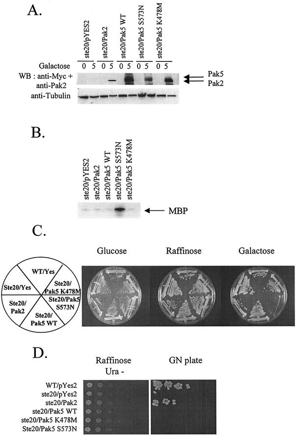
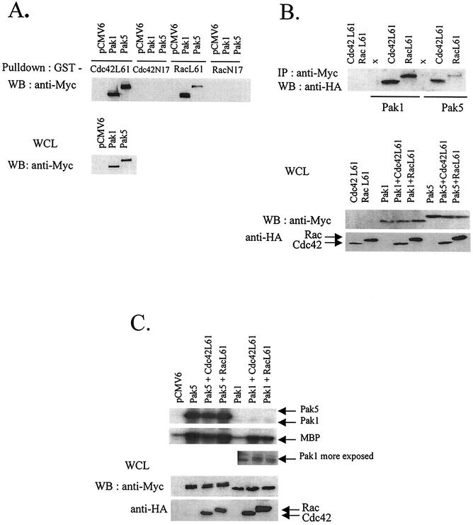
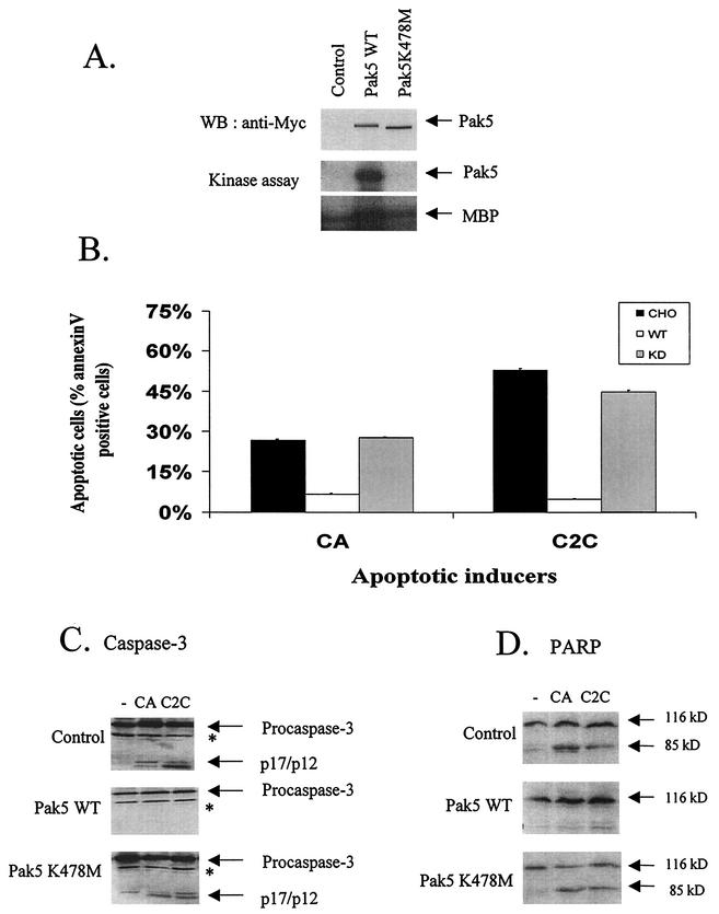
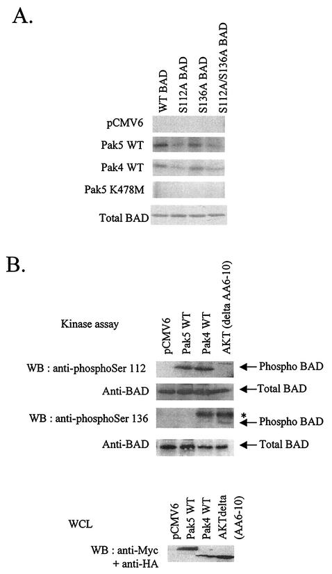
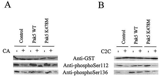
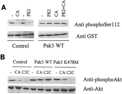
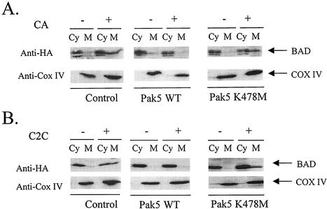
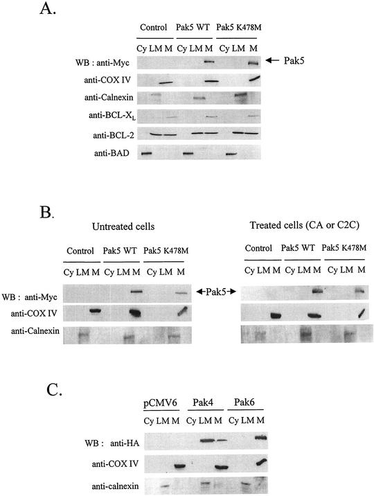
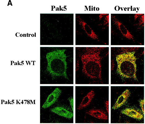
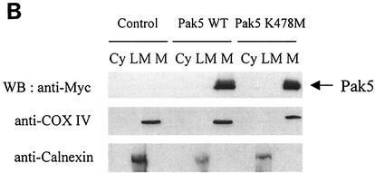
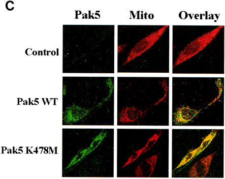
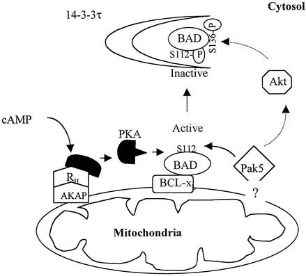
Pak5 is the most recently identified and least understood member of the p21-activated kinase (Pak) family. This kinase is known to promote neurite outgrowth in vitro, but its localization, substrates, and effects on cell survival have not been reported. We show here that Pak5 has unique properties that distinguish it from all other members of the Pak family. First, Pak5, unlike Pak1, cannot complement an STE20 mutation in Saccharomyces cerevisiae. Second, Pak5 binds to the GTPases Cdc42 and Rac, but these GTPases do not regulate Pak5 kinase activity, which is constitutive and stronger than any other Pak. Third, Pak5 prevents apoptosis induced by camptothecin and C2-ceramide by phosphorylating BAD on Ser-112 in a protein kinase A-independent manner and prevents the localization of BAD to mitochondria, thereby inhibiting the apoptotic cascade that leads to apoptosis. Finally, we show that Pak5 itself is constitutively localized to mitochondria, and that this localization is independent of kinase activity or Cdc42 binding. These features make Pak5 unique among the Pak family and suggest that it plays an important role in apoptosis through BAD phosphorylation.


Figures

FIG. 1.
FIG. 1.
Complementation analysis of Pak5. ste20-null or a control strain of S. cerevisiae was transformed with a galactose-inducible expression vector bearing either no insert or a cDNA encoding Pak2, Myc-tagged WT Pak5, hyperactivated mutant (S573N), or K478 M (KD) mutant. Transformants were selected for growth in YNB URA− raffinose medium, galactose was added to 2% at time zero, and the cultures were induced for 5 h before harvesting. (A) Expression of the transgenes. Cells were lysed before and 5 h after induction, then assayed for protein expression by immunoblotting with monoclonal anti-Myc 9E10 or anti-Pak2. Equal protein levels was assessed using an antitubulin antibody. (B) Kinase assay. Cultures tested in panel A and obtained after 5 h of induction were lysed and immunoprecipitated with a polyclonal anti-Myc antibody or polyclonal anti-Pak2 and assayed for kinase activity as described in Materials and Methods. (C) Transformants were streaked onto YNB glucose, YNB raffinose, and YNB galactose URA− plates and incubated at 30°C before being photographed. (D) To perform semiquantitative mating assays, the transformants were serially diluted onto YNB raffinose URA− plates. After the colonies had grown sufficiently, the plates were replicated onto YNB Gal URA− for 5 h to allow expression of the transgenes. The cells were then replica-plated onto YEPD plates containing a lawn of the mating tester strain, and allowed to mate for six hours before being replicated onto GN plates. Mating efficiency was scored by the formation of prototrophic strains on the GN plates.
FIG. 2.
FIG. 2.
Biochemical properties of Pak5. (A) COS cells were transfected with equal amounts of expression vectors encoding WT Myc-tagged Pak1 and Pak5 or empty vector. After transient expression, Myc-Pak1 and Myc-Pak5 were pulled down from whole-cell lysate (WCL) with GST-Cdc42 and GST-Rac loaded with GTP, Western blotting was then performed using an anti-Myc antibody. The expression level of Pak1 and Pak5 in the lysates used for the pull-down is shown in the bottom panel, expression was detected by Western blotting using an anti-Myc antibody. (B) COS cells were cotransfected with equal amounts of expression vectors encoding HA-tagged Cdc42 L61 or HA-Rac L61 or Myc-tagged Pak1 or -Pak5 alone or plus HA-tagged Cdc42 L61 or HA-Rac L61. After transient expression, Myc-Pak1 and Myc-Pak5 were immunoprecipitated from total lysates with an anti-Myc antibody and protein A-agarose. Western blotting was then performed and the complexes were revealed with an anti-HA antibody. The bottom panel shows the expression of Pak1 and Pak5, revealed with an anti-Myc and Cdc42 and Rac, revealed with an anti-HA in the WCL. (C) COS cells were transfected with expression vectors for Myc-tagged Pak1 or -Pak5 or cotransfected with Pak1 or Pak5 cDNAs and the HA-tagged GTPases Cdc42 and Rac. Myc-tagged Pak1 and Pak5 were immunoprecipitated with an anti-Myc antibody and the kinase assay was performed as described in Materials and Methods using [γ-32P]ATP and MBP as a substrate. Reaction products were separated with SDS-PAGE and subjected to autoradiography. The bottom panel shows the expression levels of proteins in the WCL prior to immunoprecipitation.
FIG. 3.
FIG. 3.
Pak5 induces resistance to apoptosis. (A) Expression and activity of Pak5 in CHO stable cell lines. Stable CHO cell lines were established to study apoptosis. Equal amounts of lysates from stable CHO cells containing empty vector, Myc-WT Pak5, or Myc-KD Pak5 were analyzed for the expression of Pak5 proteins by Western blotting using anti-Myc antibody. Kinase activity was assessed by performing a kinase assay. Myc-tagged proteins were immunoprecipitated with anti-Myc and using MBP as a substrate. (B) Stable cell lines expressing Pak5 are resistant to apoptosis. Cells were treated with C2C (50 μM) and CA (10 μM) for 16 h and were collected and stained with annexin V and propidium iodide. Apoptosis was assessed by flow cytometry. The percentages of total cells positive for annexin V are shown on the graph, and represent the average of three experiments. (C and D) Caspase 3 and PARP cleavage. Cells were treated as indicated above with C2C and CA. Cells were lysed and Western blotting was performed using anti-caspase 3 and anti-PARP antibodies. *, nonspecific band.
FIG. 4.
FIG. 4.
Pak5 phosphorylates BAD on serine 112 in vitro. (A) COS cells were transfected with empty vector or Myc-Pak5 or HA-Pak4. An equal amount of cell extracts were used for in vitro kinase assay. Myc-tagged proteins were immunoprecipitated with anti-Myc, the immunocomplexes were incubated in the presence of [γ-32P]ATP and equal amounts of recombinant His-tagged BAD (WT, S112A, S136A, and S112A/S136A) as substrates as shown on the bottom panel. After SDS-PAGE, phosphorylated BAD was detected by autoradiography. (B) The kinase assay was performed using His-tagged WT BAD, and immunocomplexes from lysates of COS cells transfected with Myc-Pak5, HA-Pak4 and HA-Akt (activated mutant, del AA6-10). After Western blotting, phosphorylated BAD was detected with anti-phospho-Ser 112 and anti-phospho-Ser 136, while total BAD was detected with an anti-BAD antibody. Bottom panel shows the expression of the different proteins in the whole-cell lysate (WCL). * shows cross-reaction of anti-phospho-Ser 136 BAD with anti-HA immunoglobulin G light chain.
FIG. 5.
FIG. 5.
Pak5 prevents BAD dephosphorylation in vivo. CHO control cells and stable cell lines expressing WT Pak5 or KD Pak5 were transfected with pEBG-BAD and were treated with CA (10 μM) or C2C (50 μM) 24 h after transfection. Equal amounts of cell extracts were analyzed by Western blotting after pull-down assay of GST-BAD, using an anti-GST antibody to assess BAD expression level, and antibodies directed against phospho-Ser 112 and phospho-Ser 136 to assess BAD phosphorylation status.
FIG. 6.
FIG. 6.
Pak5 phosphorylates BAD independently of PKA and prevents Akt dephosphorylation on serine 473. (A) Stable cell lines were transfected with pEBG-BAD and after 24 h were pretreated 30 min with a specific myristoylated protein kinase A inhibitor (1 μM) before CA (10 μM) was added for 16 h. GST-BAD was then retrieved from the lysates and an anti-phospho-Ser-112 BAD antibody was used for the Western blot to assess BAD phosphorylation status. (B) Stable cell lines were treated for 16 h with CA (10 μM) or C2C (50 μM). After lysis, Western blotting was performed using an anti-phospho-Ser 473 specific antibody.
FIG. 7.
FIG. 7.
Pak5 prevents BAD translocation to mitochondria. CHO control cells and stable cell lines expressing WT Pak5 and KD Pak5 were transfected with HA-BAD and 24 h later treated with CA or C2C for 16 h. Cells were collected by trypsinization and a cellular fractionation was performed to isolate mitochondria as described in Materials and Methods. Equal amount of proteins were separated by SDS-PAGE. BAD was detected by Western blotting using an anti-HA antibody. Equal loading of the mitochondrial pellet was assessed with an anti-COX IV antibody.
FIG. 8.
FIG. 8.
Pak5 is present in the mitochondrial fraction. A cellular fractionation was performed on CHO stable cell lines as described in Materials and Methods to assess Pak5 presence in the cellular compartments. (A) Equal amount of proteins from cytosolic (Cy) S100, light microsomes (LM) and mitochondrial (M) fractions were loaded on a gel, and Western blotting was performed using anti-Myc antibody. The mitochondrial fraction purity was assessed using an anti-COX IV and anticalnexin antibodies. The fractions were also subjected to immunoblotting with anti-BAD, anti-BCL-2 and anti-BCL-xL antibodies. (B) CHO cells expressing vector alone, WT Pak5, or KD Pak5 were treated with CA (10 μM) or C2C (50 μM) for 16 h. A cellular fractionation was then performed. Equal amount of proteins from cytosolic (Cy) S100, light microsomes (LM) and mitochondrial (M) fractions were loaded on a gel, and Western blotting was performed using anti-Myc antibody. The mitochondrial fraction purity was assessed using an anti-COX IV and anticalnexin antibodies. (C) CHO cells were transfected with pCMV6, HA-Pak4 or HA-Pak6, and a subcellular fractionation was performed 48 h after transfection as described above.
FIG. 9.
FIG. 9.
Pak5 colocalizes with mitochondria. CHO stable cell lines (A) expressing WT Pak5 or KD Pak5 were labeled as described in Materials and Methods. The fields shown were analyzed independently by digital confocal fluorescence microscopy at the appropriate wavelength for FITC (Pak5) and MitoTracker Red CMXRos (Mito), and the two images were overlaid (Overlay). (B) HMN1 cells were transfected with the empty vector (control) or with a vector carrying WT Pak5 or Pak5 K478M cDNA. A cellular fractionation was then performed 48 h after transfections as described in Materials and Methods to assess Pak5 presence in the cellular compartments. Equal amount of proteins from cytosolic (Cy) S100, light microsomes (LM) and mitochondrial (M) fractions were loaded on a gel, and Western blotting was performed using anti-Myc antibody. The mitochondrial fraction was assessed using anti-COX IV and anticalnexin antibodies. (C) HMN1 cells were transfected with empty vector or Myc-tagged WT Pak5 or KD Pak5 were stained as described in Materials and Methods. The fields shown were analyzed independently by fluorescence microscopy at the appropriate wavelength for FITC (Pak5) and MitoTracker Red CMXRos (Mito), and the two images were overlaid (Overlay).
FIG. 9.
FIG. 9.
Pak5 colocalizes with mitochondria. CHO stable cell lines (A) expressing WT Pak5 or KD Pak5 were labeled as described in Materials and Methods. The fields shown were analyzed independently by digital confocal fluorescence microscopy at the appropriate wavelength for FITC (Pak5) and MitoTracker Red CMXRos (Mito), and the two images were overlaid (Overlay). (B) HMN1 cells were transfected with the empty vector (control) or with a vector carrying WT Pak5 or Pak5 K478M cDNA. A cellular fractionation was then performed 48 h after transfections as described in Materials and Methods to assess Pak5 presence in the cellular compartments. Equal amount of proteins from cytosolic (Cy) S100, light microsomes (LM) and mitochondrial (M) fractions were loaded on a gel, and Western blotting was performed using anti-Myc antibody. The mitochondrial fraction was assessed using anti-COX IV and anticalnexin antibodies. (C) HMN1 cells were transfected with empty vector or Myc-tagged WT Pak5 or KD Pak5 were stained as described in Materials and Methods. The fields shown were analyzed independently by fluorescence microscopy at the appropriate wavelength for FITC (Pak5) and MitoTracker Red CMXRos (Mito), and the two images were overlaid (Overlay).
FIG. 10.
FIG. 10.
Model of Pak5 in cell survival signaling. Pak5 is localized to mitochondria, either directly or via interactions with a tethering protein. There, it encounters BAD, which it phosphorylates at Ser-112. The PKA complex (comprising a catalytic subunit, a regulatory subunit [RII], and a mitochondrial-tethering subunit [AKAP]) independently targets the Ser-112 site. Pak5 also induces, through an unknown mechanism, the activation of Akt, which phosphorylates BAD at Ser-136. Phosphorylated BAD complexes with 14-3-3τ in the cytosol.

Similar articles

Cited by

References

    1. Abo, A., J. Qu, M. S. Cammarano, C. Dan, A. Fritsch, V. Baud, B. Belisle, and A. Minden. 1998. PAK4, a novel effector for Cdc42Hs, is implicated in the reorganization of the actin cytoskeleton and in the formation of filopodia. EMBO J. 17:6527-6540. - PMC - PubMed
    1. Bagrodia, S., and R. A. Cerione. 1999. PAK to the future. Trends Cell Biol. 9:350-355. - PubMed
    1. Bagrodia, S., S. Taylor, C. L. Creasy, J. Chernoff, and R. A. Cerione. 1995. Identification of a murine p21Cdc42/Rac activated protein kinase (PAK). J. Biol. Chem. 270:22731-22738. - PubMed
    1. Brown, J. L., L. Stowers, M. Maer, J. Trejo, S. Coughlin, and J. Chant. 1996. Human Ste20 homologue hPak1 links GTPases to the JNK MAP kinase pathway. Curr. Biol. 6:598-605. - PubMed
    1. Cau, J., S. Faure, M. Comps, C. Delsert, and N. Morin. 2001. A novel p21-activated kinase binds the actin and microtubule networks and induces microtubule stabilization. J. Cell Biol. 155:1029-1042. - PMC - PubMed

Publication types

MeSH terms