. 2003 Aug;23(16):5664-79.
doi: 10.1128/MCB.23.16.5664-5679.2003.

BMPER, a novel endothelial cell precursor-derived protein, antagonizes bone morphogenetic protein signaling and endothelial cell differentiation

Affiliations

BMPER, a novel endothelial cell precursor-derived protein, antagonizes bone morphogenetic protein signaling and endothelial cell differentiation

Martin Moser et al. Mol Cell Biol. 2003 Aug.

Abstract

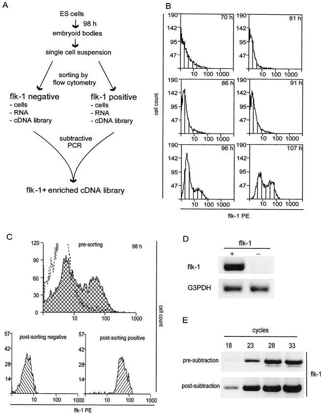
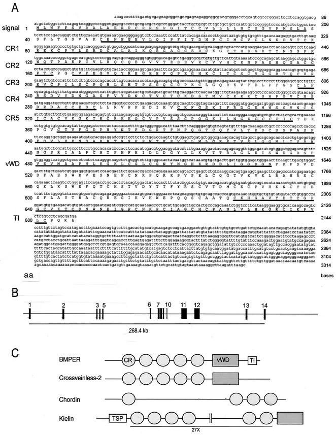
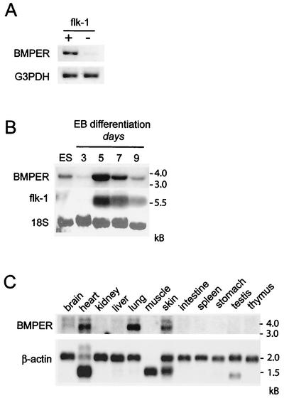
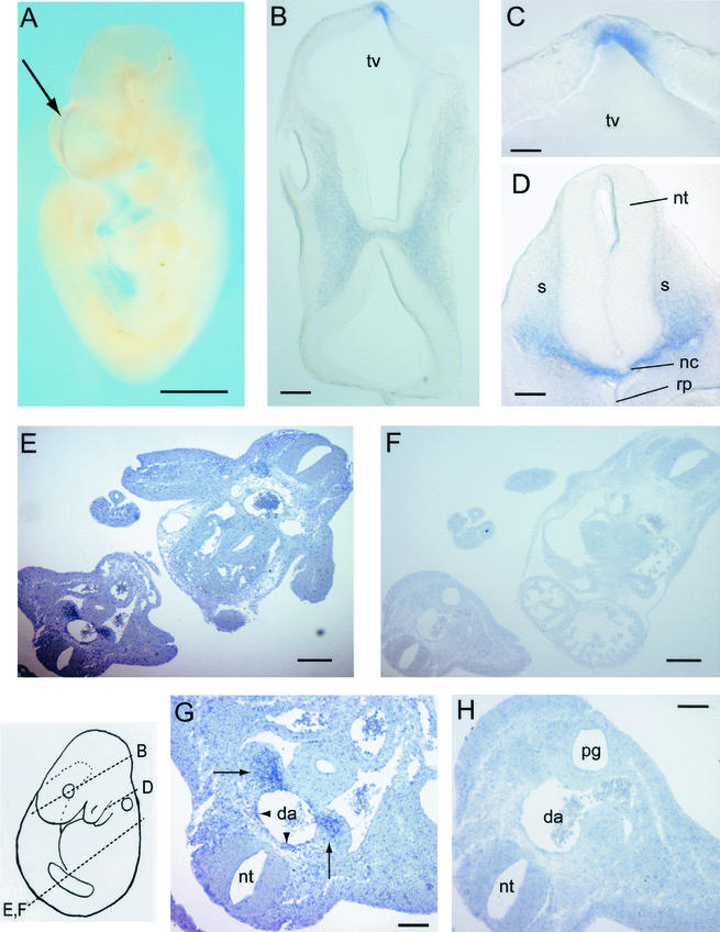
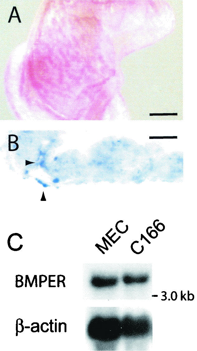
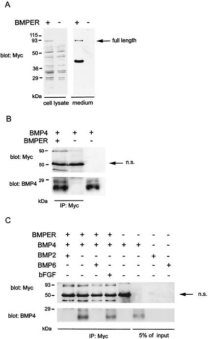
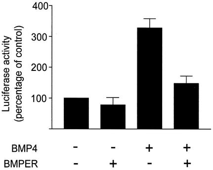
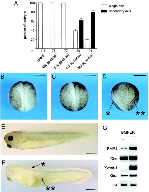
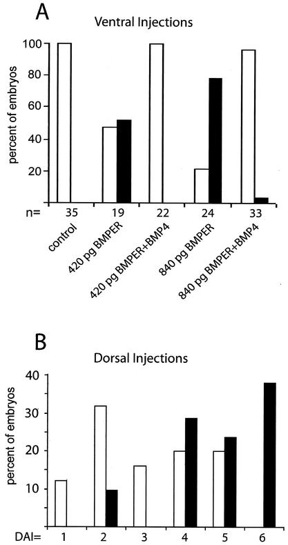
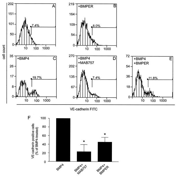
The development of endothelial cell precursors is essential for vasculogenesis. We screened for differentially expressed transcripts in endothelial cell precursors in developing mouse embryoid bodies. We cloned a complete cDNA encoding a protein that contains an amino-terminal signal peptide, five cysteine-rich domains, a von Willebrand D domain, and a trypsin inhibitor domain. We termed this protein BMPER (bone morphogenetic protein [BMP]-binding endothelial cell precursor-derived regulator). BMPER is specifically expressed in flk-1-positive cells and parallels the time course of flk-1 induction in these cells. In situ hybridization in mouse embryos demonstrates dorsal midline staining and staining of the aorto-gonadal-mesonephric region, which is known to host vascular precursor cells. BMPER is a secreted protein that directly interacts with BMP2, BMP4, and BMP6 and antagonizes BMP4-dependent Smad5 activation. In Xenopus embryos, ventral injection of BMPER mRNA results in axis duplication and downregulation of the expression of Xvent-1 (downstream target of Smad signaling). In an embryoid body differentiation assay, BMP4-dependent differentiation of endothelial cells in embryoid bodies is also antagonized by BMPER. Taken together, our data indicate that BMPER is a novel BMP-binding protein that is expressed by endothelial cell precursors, has BMP-antagonizing activity, and may play a role in endothelial cell differentiation by modulating local BMP activity.


Figures

FIG. 1.
FIG. 1.
Screening for differentially expressed cDNAs in endothelial cell precursors. (A) Outline of the screening strategy. (B) Time course of flk-1 expression in embryoid body cultures. Hours are given to indicate the time after the initiation of differentiation. Single-cell solutions of embryoid bodies were stained with PE-conjugated anti-flk-1 antibody. Subsequent studies were performed to coincide with the initial wave of flk-1 cell surface expression. (C) Separation of flk-1-positive endothelial cell precursors from flk-1-negative cells by sorting of a mixed single-cell solution of embryoid body cultures at 98 h. Reanalysis demonstrated near homogeneity of the postsorting populations. (D) PCR (25 cycles) for flk-1 with cloned cDNA libraries from flk-1-positive or -negative cells as a template. Highly efficient enrichment of flk-1 was seen. G3PDH was amplified as a control. (E) PCR for flk-1 with the flk-1-positive cDNA library before (upper panel) or after (lower panel) subtraction of the flk-1-negative library as a template. After subtraction, flk-1 was enriched approximately 32-fold (25).
FIG. 2.
FIG. 2.
Cloning and analysis of the BMPER cDNA and gene. (A) Nucleotide (upper row) and deduced amino acid (lower row) sequences. The open reading frame contains 2,055 bases coding for 685 amino acids. The predicted molecular mass is 76.1 kDa. The hydrophobic signal sequence, CR domains 1 to 5, a partial von Willebrand D (vWD) domain, and a trypsin inhibitor (TI) domain are underlined. (B) Exon-intron organization of the gene locus on mouse chromosome 9A4. (C) Structures of Crossveinless-2, Chordin, and Kielin in comparison to that of BMPER. TSP, thrombospondin domain.
FIG. 3.
FIG. 3.
BMPER mRNA expression in differentiating embryoid bodies and adult tissue. (A) Expression of BMPER in flk-1-positive and flk-1-negative cells sorted from differentiating embryoid bodies, as determined by semiquantitative PCR. (B) Northern analysis with cDNAs for BMPER, flk-1, and 18S rRNA (as an indicator of RNA loading) to probe total RNA obtained from embryoid body (EB) cultures on the indicated days of differentiation or from undifferentiated ES cells (ES). After hybridization with BMPER, a single band of 3.5 kb was visible. (C) Northern analysis with a BMPER cDNA probe of polyadenylated RNA from the indicated adult mouse tissues. Filters were probed with β-actin to visualize RNA loading. BMPER mRNA expression was highest in the heart, lungs, skin, and brain.
FIG. 4.
FIG. 4.
Localization of BMPER mRNA in developing mouse embryos. (A) Whole-mount in situ hybridization of a mouse day 9.5 embryo with a digoxigenin-labeled BMPER antisense cDNA probe. Note the midline staining in the rostral telencephalon (arrow). Bar, 500 μm. (B) Transverse section from the same embryo at the level indicated in the diagram at the lower left. Bar, 80 μm. tv, telencephalic vesicle. (C) Magnification of the frontal area from panel B. Bar, 20 μm. (D) Transverse section of the embryo shown in panel A at the level indicated in the diagram. Bar, 80 μm. nt, neural tube; rp, Rathke's pouch; s, somite; nc, notochord. Cells that appear to be migrating ventrally toward the branchial arch and heart region express BMPER. (E to H) In situ hybridization of sections from a day 10.5 mouse embryo. Arrows indicate the AGM region. (E and F) Overview of BMPER staining (E) versus that of the sense control (F) at the level indicated in the diagram. Bar, 400 μm. (G) Magnification of panel E. Bar, 150 μm. da, dorsal aorta. Note the staining of cells ventrolateral to the dorsal aorta and a ring-shaped layer of stained cells around the dorsal aorta (arrowheads). (H) Magnification of panel F. Bar, 50 μm. pg, primitive gut.
FIG. 5.
FIG. 5.
BMPER expression in extraembryonic tissues and endothelial cell lines. (A) Whole-mount in situ hybridization of a yolk sac at day 9.5. Bar, 200 μm. (B) In situ hybridization of a sectioned yolk sac at day 10.5. Arrowheads indicate BMPER-positive cells. Bar, 70 μm. (C) Northern analysis of total RNA from cultured mouse intraembryonic endothelial cells (MEC) and cultured mouse yolk sac-derived embryonic endothelial cells (C166) with a BMPER cDNA probe.
FIG. 6.
FIG. 6.
Mammalian expression of BMPER and BMP binding. (A) COS-7 cells were transiently transfected with vector pSecTag2BMPER, which is tagged with a Myc epitope at the carboxyl terminus, or empty vector. Supernatant medium and whole-cell lysates were obtained and subjected to Western blot analysis with an anti-Myc antibody. The arrow indicates full-length BMPER. (B) Myc-tagged BMPER purified from cell supernatants was incubated with recombinant BMP4, as indicated, and reaction mixtures were immunoprecipitated (IP) with an anti-Myc antibody, followed by Western blotting for BMP4 (lower panel). BMP4 was immunoprecipitated only in the presence of BMPER. In the third lane, BMP4 (10 ng) was loaded directly as a control. The efficient immunoprecipitation of BMPER was confirmed by blotting for Myc (upper panel). n.s., nonspecific. (C) Competition with BMP4 binding was used to determine the promiscuity of BMPER interactions with BMPs. Competition for coimmunoprecipitation of BMP4 with BMPER was tested in the presence of a 25-fold molar excess of BMP2, BMP6, or bovine fibroblast growth factor (bFGF).
FIG. 7.
FIG. 7.
Effects of BMPER expression on BMP4 activity in mammalian cells. 293T cells were transiently transfected with an Smad5-dependent luciferase reporter plasmid and pSecTag2BMPER or control plasmid, as indicated. A plasmid expressing β-galactosidase was cotransfected to control for transfection efficiency. Recombinant BMP4 or a control, as indicated, was added after 18 h and incubated for an additional 20 h. Luciferase activity was normalized for transfection efficiency and expressed as a percentage of the activity in untreated cells (mean and standard deviation).
FIG. 8.
FIG. 8.
Effects of BMPER on dorsal-ventral axis development in X. laevis. (A) Summary of the effects of BMPER injection into Xenopus blastomeres at the four-cell stage. n, number of embryos. Error bars indicate standard deviations. (B to D) Axis formation in Xenopus embryos at stage 14. Bar, 750 μm. (B) Control. (C) Dorsally injected. (D) Ventrally injected. Single asterisk, primary axis; double asterisk, secondary axis. (E and F) Representative embryos at stage 32. Bar, 1 mm. (E) Control. (F) Ventrally injected. (G) Mesoderm marker gene expression in activin A-treated animal caps, as analyzed by RT-PCR at stage 10. +, injected; −, not injected; H4, histone H4.
FIG. 9.
FIG. 9.
Coinjection of BMPER and BMP4. (A) Effects of coinjection of BMPER and BMP4 (50 pg) into both ventral blastomeres at the four-cell stage versus injection of BMPER alone. Embryos were scored for secondary axis formation at stage 14. Symbols: □, single axis; ▪, secondary axis. (B) Effects of coinjection of BMPER (840 pg) and BMP4 (50 pg) into both dorsal blastomeres versus injection of BMP4 alone. Embryos were scored for dorsoanterior structures at stage 32. DAI, dorsoanterior index. Symbols: □, BMP4, 25 embryos; ▪, BMP4 plus BMPER, 21 embryos.
FIG. 10.
FIG. 10.
Effects of BMPER on BMP-dependent differentiation of ES cells into endothelial cells. Embryoid bodies were incubated for 7 days under serum-free conditions. Definitive endothelial cells were detected by flow cytometry with an anti-VE-cadherin rat immunoglobulin G2 antibody and the respective fluorescein isothiocyanate (FITC)-conjugated secondary antibody. (A and B) VE-cadherin-positive cells were rare in the absence of BMP4 (A), and BMPER expression had a minimal effect under these conditions (B). (C and D) Expansion of the endothelial cell population increased in the presence of recombinant BMP4 (1 nM) (C), but this effect was attenuated when recombinant BMP4 was preincubated for 30 min at 37°C with a blocking antibody (MAB757) at a final concentration of 2 μg/ml (D). (E) Similarly, the expression of BMPER inhibited endothelial cell differentiation induced by BMP4. Results for panels A to E are representative of three independent experiments. (F) BMP4-dependent endothelial cell differentiation in embryoid bodies, as determined on the basis of VE-cadherin-positive cells in treated cells. Error bars indicate standard errors of the mean. An asterisk indicates a P value of <0.05 for a comparison with BMP4-treated cells.

Similar articles

Cited by

References

    1. Abreu, J. G., N. I. Ketpura, B. Reversade, and E. M. De Robertis. 2002. Connective-tissue growth factor (CTGF) modulates cell signalling by BMP and TGF-beta. Nat. Cell Biol. 4:599-604. - PMC - PubMed
    1. Adelman, C. A., S. Chattopadhyay, and J. J. Bieker. 2002. The BMP/BMPR/Smad pathway directs expression of the erythroid-specific EKLF and GATA1 transcription factors during embryoid body differentiation in serum-free media. Development 129:539-549. - PubMed
    1. Allen, S., A. M. Abuzenadah, J. L. Blagg, J. Hinks, I. M. Nesbitt, A. C. Goodeve, T. Gursel, J. Ingerslev, I. R. Peake, and M. E. Daly. 2000. Two novel type 2N von Willebrand disease-causing mutations that result in defective factor VIII binding, multimerization, and secretion of von Willebrand factor. Blood 95:2000-2007. - PubMed
    1. Balemans, W., and W. vanHul. 2002. Extracellular regulation of BMP signaling in vertebrates: a cocktail of modulators. Dev. Biol. 250:231-250. - PubMed
    1. Bautch, V. 2002. Embryonic stem cell differentiation and the vascular lineage. Humana Press, Totowa, N.J. - PubMed

Publication types

MeSH terms

Substances