. 2003 Aug;23(16):5706-15.
doi: 10.1128/MCB.23.16.5706-5715.2003.

Pathways of DNA double-strand break repair during the mammalian cell cycle

Affiliations

Pathways of DNA double-strand break repair during the mammalian cell cycle

Kai Rothkamm et al. Mol Cell Biol. 2003 Aug.

Abstract

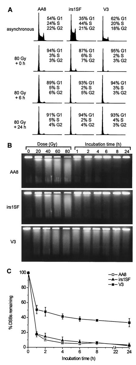
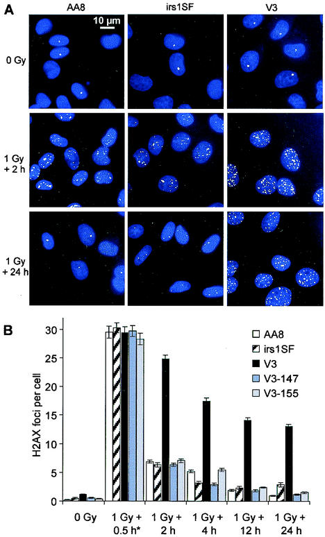
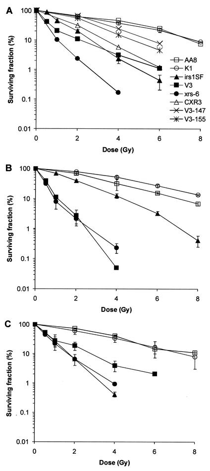
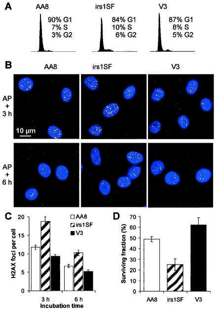
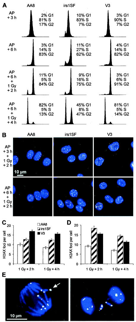
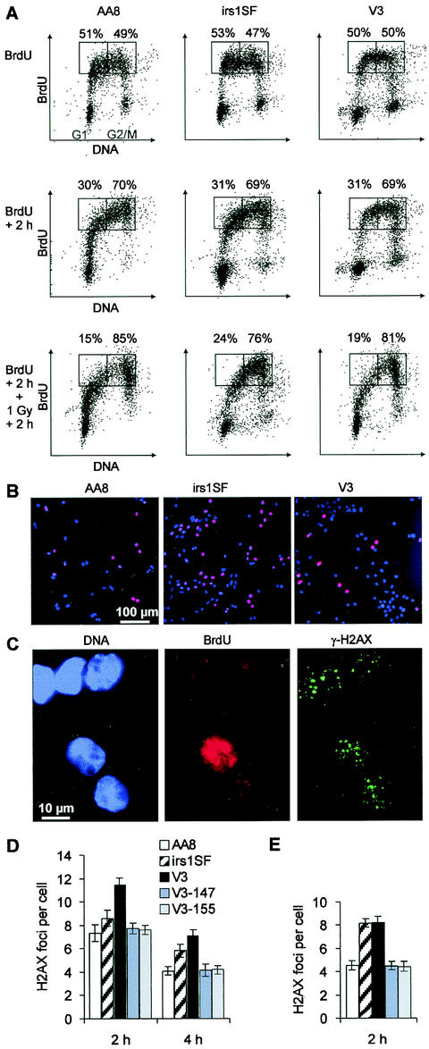
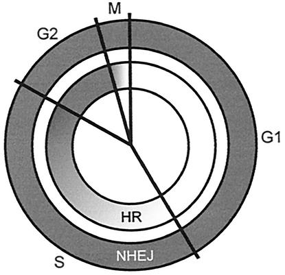
Little is known about the quantitative contributions of nonhomologous end joining (NHEJ) and homologous recombination (HR) to DNA double-strand break (DSB) repair in different cell cycle phases after physiologically relevant doses of ionizing radiation. Using immunofluorescence detection of gamma-H2AX nuclear foci as a novel approach for monitoring the repair of DSBs, we show here that NHEJ-defective hamster cells (CHO mutant V3 cells) have strongly reduced repair in all cell cycle phases after 1 Gy of irradiation. In contrast, HR-defective CHO irs1SF cells have a minor repair defect in G(1), greater impairment in S, and a substantial defect in late S/G(2). Furthermore, the radiosensitivity of irs1SF cells is slight in G(1) but dramatically higher in late S/G(2), while V3 cells show high sensitivity throughout the cell cycle. These findings show that NHEJ is important in all cell cycle phases, while HR is particularly important in late S/G(2), where both pathways contribute to repair and radioresistance. In contrast to DSBs produced by ionizing radiation, DSBs produced by the replication inhibitor aphidicolin are repaired entirely by HR. irs1SF, but not V3, cells show hypersensitivity to aphidicolin treatment. These data provide the first evaluation of the cell cycle-specific contributions of NHEJ and HR to the repair of radiation-induced versus replication-associated DSBs.


Figures

FIG. 1.
FIG. 1.
DSB repair in G1-phase cells, as determined by PFGE. (A) DNA histograms for asynchronous cultures (upper row) and G1-phase cells at various incubation times after 80 Gy of irradiation (lower rows). (B) Ethidium bromide images of DNA from G1-phase cells irradiated with various doses and incubated for repair after 80 Gy of irradiation. (C) Time course for the percentage of initial DSBs remaining after repair. Error bars represent SEMs from two or three experiments.
FIG. 2.
FIG. 2.
DSB repair in G1-phase cells, as measured by γ-H2AX focus formation. (A) γ-H2AX foci (green) in nonirradiated and irradiated cells; nuclei were stained with 4,6-diamidino-2-phenylindole (blue). (B) Time course for the repair of DSBs after 1 Gy of irradiation. The mean number of foci per cell for various repair times is shown. Numbers of foci in nonirradiated controls were subtracted. The 0.5-h time point (asterisk) was obtained by incubating the cells in the presence of LY294002 to inhibit repair. Error bars represent SEMs.
FIG. 3.
FIG. 3.
Radiosensitivity of various CHO cell lines. xrs-6 is defective in Ku80 and is derived from CHO K1 cells. (A) Asynchronous cells. (B) G1 phase. (C) Late S/G2 phase. Error bars represent SEMs from one or two (A) or two or three (B and C) independent experiments.
FIG. 4.
FIG. 4.
Effect of aphidicolin on cell cycle arrest, γ-H2AX focus formation, and cell survival. (A) DNA histograms for cells treated for 16 h with aphidicolin. (B) γ-H2AX foci (green) 3 and 6 h (3.5 and 6.5 h for irs1SF cells because of their slower growth) after treatment with aphidicolin (AP); nuclei were stained with 4,6-diamidino-2-phenylindole (blue). (C) Mean number of foci per cell after 3 and 6 h (3.5 and 6.5 h for irs1SF cells) after aphidicolin treatment. (D) Cell survival after aphidicolin treatment. Error bars represent SEMs.
FIG. 5.
FIG. 5.
DSB repair in S and G2-phase cells, as measured by γ-H2AX focus formation. (A) DNA histograms either 3 h (first row; 3.5 h for irs1SF cells) and 6 h (second row; 6.5 h for irs1SF cells) after aphidicolin (AP) removal or 2 h (third row) and 4 h (fourth row) after 1 Gy of irradiation given at 6 or 6.5 h after aphidicolin removal. (B) γ-H2AX foci (green) 2 h after 1 Gy of irradiation at 3 or 3.5 h (upper row) and 6 or 6.5 h (lower row) after aphidicolin removal; nuclei were stained with 4,6-diamidino-2-phenylindole (blue). (C) Mean number of foci per cell 2 and 4 h after 1 Gy of irradiation at 3 or 3.5 h after aphidicolin removal. (D) Mean number of foci per cell 2 and 4 h after 1 Gy of irradiation at 6 or 6.5 h after aphidicolin removal. Error bars represent SEMs. (E) γ-H2AX foci (green) in mitotic AA8 cells be-tween 2 and 4 h after 1 Gy of irradiation at 6 h after aphidicolin removal; DNA was stained with 4,6-diamidino-2-phenylindole (blue). The γ-H2AX focus indicated by the arrow coincides with a small chromosomal fragment that was invisible due to the superposition of the blue and the green images.
FIG. 6.
FIG. 6.
DSB repair in BrdU-positive cells, as measured by γ-H2AX focus formation. (A) Two-parameter (propidium iodide and BrdU) flow cytometry diagrams of asynchronous cultures of AA8, irs1SF, and V3 cells either immediately (upper row) or 2 h (middle row) after 30 min of pulse-labeling with BrdU or 2 h after 1 Gy of irradiation at 2 h after BrdU labeling (lower row). The numbers above the rectangles in each diagram refer to the proportions of BrdU-positive cells with a DNA content either lower or higher than that of mid-S-phase cells. (B) Asynchronous cultures of AA8, irs1SF, and V3 cells after 30 min of pulse-labeling with BrdU (red); nuclei were stained with 4,6-diamidino-2-phenylindole (blue). (C) γ-H2AX foci (green) in AA8 cells 2 h after 1 Gy of irradiation immediately after BrdU labeling (red); nuclei were stained with 4,6-diamidino-2-phenylindole (blue). (D) Meannumber of foci per BrdU-positive cell 2 and 4 h after 1 Gy of irradiation immediately after BrdU labeling. (E) Mean number of foci per BrdU-positive cell 2 h after 1 Gy of irradiation at 2 h after BrdU labeling. Numbers of foci in nonirradiated controls were subtracted. Error bars represent SEMs. Results obtained from a second independent set of experiments with AA8, irs1SF, and V3 cells were nearly identical to the data shown in panels D and E.
FIG. 7.
FIG. 7.
Model of the relative contributions of HR and NHEJ to the repair of IR-induced DSBs in different cell cycle phases, based on mutant phenotypes. Whereas NHEJ predominates in G1/early S, both HR and NHEJ contribute substantially to DSB repair during late S/G2.

Similar articles

Cited by

References

    1. Arnaudeau, C., C. Lundin, and T. Helleday. 2001. DNA double-strand breaks associated with replication forks are predominantly repaired by homologous recombination involving an exchange mechanism in mammalian cells. J. Mol. Biol. 307:1235-1245. - PubMed
    1. Asaad, N. A., Z. C. Zeng, J. Guan, J. Thacker, and G. Iliakis. 2000. Homologous recombination as a potential target for caffeine radiosensitization in mammalian cells: reduced caffeine radiosensitization in XRCC2 and XRCC3 mutants. Oncogene 19:5788-5800. - PubMed
    1. Baumann, P., and S. C. West. 1997. The human Rad51 protein: polarity of strand transfer and stimulation by hRP-A. EMBO J. 16:5198-5206. - PMC - PubMed
    1. Baumann, P., and S. C. West. 1998. Role of the human RAD51 protein in homologous recombination and double-stranded-break repair. Trends Biochem. Sci. 23:247-251. - PubMed
    1. Bezzubova, O., A. Silbergleit, Y. Yamaguchi-Iwai, S. Takeda, and J. M. Buerstedde. 1997. Reduced X-ray resistance and homologous recombination frequencies in a RAD54−/− mutant of the chicken DT40 cell line. Cell 89:185-193. - PubMed

Publication types

Substances

LinkOut - more resources