. 2003 Aug;23(16):5836-48.
doi: 10.1128/MCB.23.16.5836-5848.2003.

Autophosphorylation of the catalytic subunit of the DNA-dependent protein kinase is required for efficient end processing during DNA double-strand break repair

Affiliations

Autophosphorylation of the catalytic subunit of the DNA-dependent protein kinase is required for efficient end processing during DNA double-strand break repair

Qi Ding et al. Mol Cell Biol. 2003 Aug.

Abstract

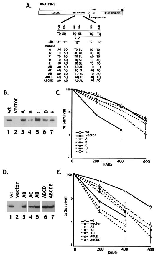
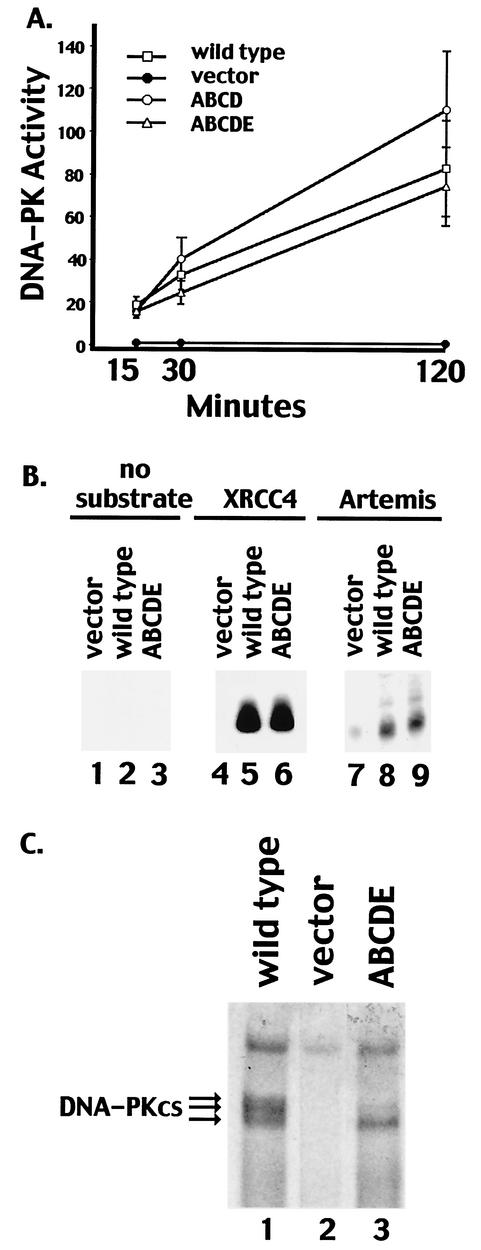
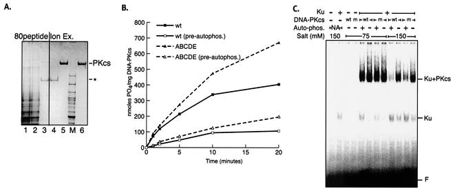
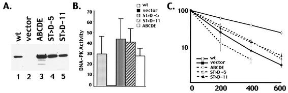
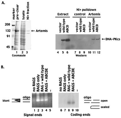
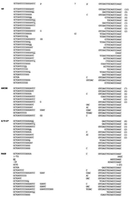
The DNA-dependent protein kinase (DNA-PK) plays an essential role in nonhomologous DNA end joining (NHEJ) by initially recognizing and binding to DNA breaks. We have shown that in vitro, purified DNA-PK undergoes autophosphorylation, resulting in loss of activity and disassembly of the kinase complex. Thus, we have suggested that autophosphorylation of the DNA-PK catalytic subunit (DNA-PKcs) may be critical for subsequent steps in DNA repair. Recently, we defined seven autophosphorylation sites within DNA-PKcs. Six of these are tightly clustered within 38 residues of the 4,127-residue protein. Here, we show that while phosphorylation at any single site within the major cluster is not critical for DNA-PK's function in vivo, mutation of several sites abolishes the ability of DNA-PK to function in NHEJ. This is not due to general defects in DNA-PK activity, as studies of the mutant protein indicate that its kinase activity and ability to form a complex with DNA-bound Ku remain largely unchanged. However, analysis of rare coding joints and ends demonstrates that nucleolytic end processing is dramatically reduced in joints mediated by the mutant DNA-PKcs. We therefore suggest that autophosphorylation within the major cluster mediates a conformational change in the DNA-PK complex that is critical for DNA end processing. However, autophosphorylation at these sites may not be sufficient for kinase disassembly.


Figures

FIG. 1.
FIG. 1.
Single phosphorylation site mutants complement the radiosensitivity of V3 cells, whereas multiple phosphorylation site mutants do not. (A) Diagrammatic representation of seven autophosphorylation sites within DNA-PKcs (asterisks). Mutations are as follows: A, T2609A; B, T2620A and S2624A; C, T2638A; D, T2647A; E, S2612A. The A, B, C, D, and E mutations were introduced either alone or in combinations as indicated. (B) Immunoblot analyses of whole-cell extracts from V3 transfectants expressing either full-length DNA-PKcs (lane 1), vector alone (lane 2), mutant A (lane 3), mutant B (lane 4), mutant C (lane 5), mutant D (lane 6), or mutant E (lane 7). (C) The radiation resistance of V3 transfectants expressing wild-type DNA-PKcs, vector alone, or each of the single phosphorylation site mutants was assessed as described in Materials and Methods. Data are presented as percent survival relative to that of nonirradiated controls (set at 100%). Error bars, standard errors of the means of three separate experiments. (D) Immunoblot analyses of whole-cell extracts from V3 transfectants expressing either full-length DNA-PKcs (lane 1), vector alone (lane 2), mutant AB (lane 3), mutant AC (lane 4), mutant AD (lane 5), mutant ABCD (lane 6), or mutant ABCDE (lane 7). (E) The radiation resistance of V3 transfectants expressing either wild-type DNA-PKcs, vector alone, or mutant AB, AC, AD, ABCD, or ABCDE was assessed. Data are presented as percent survival relative to that of nonirradiated controls (set at 100%). Error bars, standard errors of the means of three separate experiments.
FIG. 2.
FIG. 2.
Transfectants expressing multiple phosphorylation site substitutions express normal levels of DNA-PK activity. (A) Whole-cell extracts (500 μg) prepared from V3 cells transfected with vector alone, wild-type DNA-PKcs, mutant ABCD, or mutant ABCDE were assayed for DNA-PK activity as described in Materials and Methods. Phosphorylation of the p53 substrate was assessed after 15, 30, and 120 min as indicated. Each cell extract was tested in duplicate, and three independent extracts were tested for each cell line. Error bars, standard deviations. (B) Whole-cell extracts (250 μg) from V3 cells transfected with vector alone, wild-type DNA-PKcs, or mutant ABCDE were assayed for the capacity to phosphorylate no substrate (lanes 1 to 3), recombinant XRCC4 (lanes 4 to 6), or recombinant Artemis (lanes 7 to 9). (C) DNA-cellulose fractions from 500 μg of whole-cell extracts of V3 cells expressing wild-type DNA-PKcs (lane 1), vector alone (lane 2), or mutant ABCDE (lane 3) were incubated in protein kinase buffer with [γ-32P]ATP. Subsequently, bound proteins were analyzed by SDS-4% PAGE and autoradiography.
FIG. 3.
FIG. 3.
Autophosphorylation within the 2609-to-2647 cluster is not required for autophosphorylation-induced kinase dissociation. (A) Wild-type and ABCDE mutant DNA-PKcs's were purified as described in Materials and Methods. Coomassie-stained SDS-PAGE gel shows analysis of fractions from a purification of the recombinant ABCDE mutant of DNA-PKcs. Lane 1, 10 μg of clarified extract from V3 cells expressing the ABCDE mutant; lane 2, 10 μg of flowthrough after the extract was loaded onto an affinity column coupled with a peptide from the C terminus of Ku80 (80peptide); lane 3, 2 μg of pooled fractions containing DNA-PKcs from 80peptide eluate; lane 4, 2 μg of flowthrough of Mono S column; lane 5, 2 μg of pooled DNA-PKcs-containing fractions from Mono S; lane 6, 2 μg of purified DNA-PKcs from placenta. Asterisk indicates location of major contaminant in eluate from 80peptide column. Similar results were obtained by purifying recombinant wild-type DNA-PKcs. (B) Kinase activities of purified wild-type and ABCDE mutant DNA-PKcs's were assessed as described in Materials and Methods. DNA-protein complexes were preformed by incubation (at 30°C for 10 min) of 1 nM purified recombinant Ku, 1 nM either purified wild-type (wt) or purified ABCDE mutant DNA-PKcs, and 10 μg of sheared calf thymus DNA/ml. Reactions were started by transfer to 37°C and addition of 400 μM peptide substrate, 200 μM [γ-32P]ATP (0.5 Ci/mmol), and 5 mM MgCl2. The impact of prior autophosphorylation (pre-autophos.) was assessed in parallel reactions by including the same concentrations of ATP and MgCl2 in the preincubation step as well. (C) The stabilities of DNA-PK complexes containing wild-type or mutant DNA-PKcs were assessed by EMSA as described in Materials and Methods. DNA-protein complexes were formed with a 60-bp radiolabeled duplex, Ku, and wt or ABCDE mutant (m) DNA-PKcs as indicated. Complexes were preincubated at 75 mM salt by using either mock-treated DNA-PKcs, active kinase (+ Auto-phos.), or wortmannin-inactivated kinase (− Auto-phos.). Autophosphorylation was arrested by addition of 5 mM EDTA. Reactions were then adjusted to the indicated salt concentrations and incubated for a further 10 min at room temperature before surviving DNA-protein complexes were fixed by cross-linking with 0.25% glutaraldehyde. DNA-protein complexes were resolved by electrophoresis on a 3.5% native polyacrylamide gel.
FIG. 4.
FIG. 4.
An ABCDE mutant with aspartic acid substitutions is partially competent in NHEJ. (A) Immunoblot analyses of whole-cell extracts from V3 transfectants expressing wild-type (wt) DNA-PKcs (lane 1), vector alone (lane 2), mutant ABCDE (lane 3), the ST→D mutant (clone 5), (lane 4), and the ST→D mutant (clone 11) (lane 5). (B) Whole-cell extracts (500 μg) prepared from V3 cells transfected with either vector alone, wt DNA-PKcs, mutant ABCDE, or one of two independent clones (clones 5 and 11) expressing the ST→D mutation were assayed for DNA-PK activity. Phosphorylation of the p53 substrate was assessed after 60 min. Each cell extract was tested in duplicate, and three independent extracts were tested for each cell line. Error bars, standard deviations. (C) Radiation resistance of V3 transfectants expressing wt DNA-PKcs, vector alone, mutant ABCDE, or the ST→D mutant. Data are presented as percent survival relative to that of nonirradiated controls (set at 100%). Error bars, standard errors of the means of three separate experiments.
FIG. 5.
FIG. 5.
Autophosphorylation within the 2609-to-2647 cluster is not required for the interaction of DNA-PKcs with Artemis or to facilitate opening of coding end hairpins. (A) (Left) SDS-PAGE analyses of the following fractions of Sf9 cells infected with a baculovirus encoding an Artemis-V5 His-tagged fusion protein: precleared whole-cell lysate (lane 1) and cleared lysate (lane 2). Marker, marker proteins, with molecular weights (in thousands) given on the left. (Right) Western blot of whole-cell extracts or Ni+-agarose fractions. Lanes 4 to 6, whole-cell extracts of V3 cells transfected with vector alone (lane 4), wild-type DNA-PKcs (lane 5), or mutant ABCD (lane 6); lanes 7 to 9, Ni+-agarose fractions of control Sf9 extracts coincubated with extracts from V3 cells transfected with vector alone (lane 7), wild-type DNA-PKcs (lane 8), or mutant ABCD (lane 9); lanes 10 to 12, Ni+-agarose fractions of extracts from Sf9 cells expressing Artemis coincubated with extracts from V3 cells transfected with vector alone (lane 10), wild-type DNA-PKcs (lane 11), or mutant ABCD (lane 12). (B) LMPCR was performed on Hirt supernatants prepared from V3 cells transiently transfected with substrate alone (lanes 1 and 6), substrate and RAG expression vectors (lanes 2 and 7), substrate plus RAG and wild-type DNA-PKcs expression vectors (lanes 3 and 8), or substrate plus RAG and mutant ABCDE expression vectors (lanes 4 and 9). Lanes 5 and 10 included no input DNA in the LMPCRs. In lanes 1 to 5, a blunt oligonucleotide was ligated to Hirt fractions, and PCR amplifications to detect signal ends were performed. In lanes 6 to 10, an oligonucleotide with a 4-bp overhang complementary to a potential opened coding end in substrate pJH290 was ligated to Hirt fractions, and PCR amplifications to detect coding ends were performed.
FIG. 6.
FIG. 6.
Coding joints mediated by mutant ABCDE have minimal nucleotide loss from joined ends. The sequences of coding ends as they occur in the pJH290 substrate are shown above the sequences of the recombinant junctions. The number of observations of each sequence is given in parentheses to the right of the sequence. All duplicate sequences were derived from separate transfections. Nucleotides in columns headed by “p” are palindromic nucleotides added to each junction. Nucleotides that cannot be unequivocally assigned to a particular coding end are underlined and listed in the 5′-most location. Portions of the wild-type (WT) and RAG-only coding joints are from references and .

Similar articles

Cited by

References

    1. Baumann, P., and S. C. West. 1998. DNA end-joining catalyzed by human cell-free extracts. Proc. Natl. Acad. Sci. USA 95:14066-14070. - PMC - PubMed
    1. Bogue, M. A., C. Jhappan, and D. B. Roth. 1998. Analysis of variable (diversity) joining recombination in DNA-dependent protein kinase (DNA-PK)-deficient mice reveals DNA-PK-independent pathways for both signal and coding joint formation. Proc. Natl. Acad. Sci. USA 95:15559-15564. - PMC - PubMed
    1. Chan, D. W., B. P. Chen, S. Prithivirajsingh, A. Kurimasa, M. D. Story, J. Qin, and D. J. Chen. 2002. Autophosphorylation of the DNA-dependent protein kinase catalytic subunit is required for rejoining of DNA double-strand breaks. Genes Dev. 16:2333-2338. - PMC - PubMed
    1. Chan, D. W., and S. P. Lees-Miller. 1996. The DNA-dependent protein kinase is inactivated by autophosphorylation of the catalytic subunit. J. Biol. Chem. 271:8936-8941. - PubMed
    1. Chan, D. W., R. Ye, C. J. Veillette, and S. P. Lees-Miller. 1999. DNA-dependent protein kinase phosphorylation sites in Ku 70/80 heterodimer. Biochemistry 38:1819-1828. - PubMed

Publication types

MeSH terms