. 2003 Aug;23(16):5939-46.
doi: 10.1128/MCB.23.16.5939-5946.2003.

Replication checkpoint kinase Cds1 regulates recombinational repair protein Rad60

Affiliations

Replication checkpoint kinase Cds1 regulates recombinational repair protein Rad60

Michael N Boddy et al. Mol Cell Biol. 2003 Aug.

Abstract

Genome integrity is protected by Cds1 (Chk2), a checkpoint kinase that stabilizes arrested replication forks. How Cds1 accomplishes this task is unknown. We report that Cds1 interacts with Rad60, a protein required for recombinational repair in fission yeast. Cds1 activation triggers Rad60 phosphorylation and nuclear delocalization. A Rad60 mutant that inhibits regulation by Cds1 renders cells specifically sensitive to replication fork arrest. Genetic and biochemical studies indicate that Rad60 functions codependently with Smc5 and Smc6, subunits of an SMC (structural maintenance of chromosomes) complex required for recombinational repair. These studies indicate that regulation of Rad60 is an important part of the replication checkpoint response controlled by Cds1. We propose that control of Rad60 regulates recombination events at stalled forks.


Figures
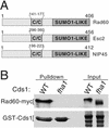
FIG. 1.
FIG. 1.
Rad60 associates with Cds1. (A) Members of the Rad60 family. Shown are Rad60 of fission yeast (406 amino acids), Esc2 of budding yeast (456 amino acids), and Nip45 of humans (412 amino acids). The C terminus of each protein contains a ubiquitin-like domain related to SUMO-1 (none has the C-terminal motifs for covalent attachment to other proteins). All contain a central coiled-coil domain (C/C). (B) Confirmation of the Rad60-Cds1 interaction in vivo. GST fusions of wild-type (WT) or mutant Cds1 (fha1) were expressed in cells that express Rad60-myc from the rad60 genomic locus. Rad60-myc coprecipitates with the wild-type but not mutant Cds1 (fha1). Approximately 1% of the total Rad60-myc coprecipitated with GST-Cds1.
FIG. 2.
FIG. 2.
Cds1 controls Rad60 phosphorylation. (A) Wild-type (WT), cds1-fha1, and cds1Δ cells were treated or left untreated with 12 mM HU for 4 h. The electrophoretic mobility of Rad60-myc was analyzed in each strain. HU caused the appearance of a reduced-mobility form of Rad60-myc in wild-type but not cds1-fha1 and cds1Δ cells. (B) Precipitates of Rad60-TAP from wild-type cells, treated or not with HU, were subjected to lambda phosphatase treatment. The slow-migrating forms of Rad60-TAP induced by HU were converted to a single faster-migrating species, showing that Rad60 is phosphorylated in response to HU treatment. The phosphatase inhibitor vanadate largely blocks the conversion of Rad60-TAP to the high-mobility species.
FIG. 3.
FIG. 3.
Mutant rad60-3 cells are hypersensitive to HU. (A) The indicated strains were serially diluted (∼2,500, 500, 100, and 20 cells per spot) and plated on medium supplemented or not with 5 mM HU followed by incubation at 25°C. (B) Cells of the indicated strains were photographed on agar medium supplemented with 5 mM HU.
FIG. 4.
FIG. 4.
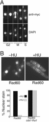
Cds1 controls nuclear delocalization of Rad60 in HU-treated cells. (A) Rad60 is a nuclear protein throughout the cell cycle. The localization of endogenous Rad60 was determined by indirect immunofluorescence of 13myc-tagged protein. The Rad60-myc signal was strongest in the chromatin (DAPI staining) region of the nucleus. Cell cycle position was determined by DAPI stain and by a phase-contrast photo (data not shown) showing whether a septum was present in binucleate cells. (B) Rad60-myc delocalizes from the nucleus during replication arrest. Rad60-myc cells were treated or not with 10 mM HU for 4 h and fixed, and Rad60-myc was detected by indirect immunofluorescence. The percentage of cells with exclusively nuclear staining was determined and is shown at the bottom. Rad60-myc delocalization was abrogated by the cds1-fha1 mutation (right panel).
FIG. 5.
FIG. 5.
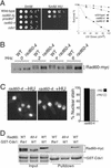
A rad60 mutant insensitive to control by Cds1. (A) Mutant rad60-4 cells are sensitive to HU but relatively insensitive to UV. Cells were serially diluted (∼2,500, 500, 100, and 20 cells per spot) and plated in the presence or absence of 5 mM HU (left panel). A single integrated copy of wild-type rad60+ (prad60+) allowed rad60-4 cells to form colonies in the presence of 5 mM HU. Survival analysis of wild-type, rad60-3, and rad60-4 cells irradiated with UV is shown in the right panel. Cells were maintained at 25°C. (B) Electrophoretic retardation of Rad60 that induced by HU was largely eliminated in rad60-4 cells. Rad60-myc (wild type [WT]) and rad60-4-myc (rad60-4) cells were treated with 12 mM HU from 0 to 6 h. Samples were taken every 2 h and analyzed by SDS-PAGE. (C) Nuclear delocalization of Rad60 induced by 10 mM HU at 4 h was abrogated in rad60-4 cells. (D) The interaction between Rad60 and Cds1 was abolished by rad60-4. GST fusions of wild-type or mutant Cds1 (fha1) were expressed in cells that express myc epitope-tagged wild type Rad60 or rad60-4 from the rad60 genomic locus. Wild-type Rad60 coprecipitated with GST-Cds1 but not GST-Cds1-fha1. Rad60-4 did not coprecipitate with GST-Cds1. In addition, Rad60 but not Rad60-4 displayed maximum electrophoretic retardation caused by phosphorylation in response to expression of GST-Cds1 but not GST-Cds1-fha1.
FIG. 6.
FIG. 6.
Rad60 interacts with Smc5 and Smc6. (A) Smc5 peptides identified in Rad60-TAP preparation. (B) GST or GST-Rad60 were expressed in strains that expressed 13myc-tagged Smc5 (Spr18) or 3myc-tagged Smc6 (Rad18) from their genomic loci (left and right panels, respectively). GST and GST-Rad60 were purified, and coprecipitating proteins were analyzed by anti-myc immunoblot (upper panels). The lower panels show Coomassie blue staining of purified GST-Rad60 and GST. Smc5 and Smc6 coprecipitated with GST-Rad60 but not GST. (C) Summary of synthetic lethal interactions involving rad60 and other genes. Double-headed arrows indicate synthetic lethality. The right-hand panel shows the rad60-3 mus81 synthetic lethal phenotype, following germination and formation of an approximately five-cell colony. Shown are cells on a tetrad dissection plate; wild-type cells had formed large colonies (data not shown).

Similar articles

Cited by

References

    1. Aono, N., T. Sutani, T. Tomonaga, S. Mochida, and M. Yanagida. 2002. Cnd2 has dual roles in mitotic condensation and interphase. Nature 417:197-202. - PubMed
    1. Bashkirov, V. I., J. S. King, E. V. Bashkirova, J. Schmuckli-Maurer, and W.-D. Heyer. 2000. DNA repair protein Rad55 is a terminal substrate of the DNA damage checkpoints. Mol. Cell. Biol. 20:4393-4404. - PMC - PubMed
    1. Birkenbihl, R. P., and S. Subramani. 1995. The rad21 gene product of Schizosaccharomyces pombe is a nuclear, cell cycle-regulated phosphoprotein. J. Biol. Chem. 270:7703-7711. - PubMed
    1. Boddy, M. N., B. Furnari, O. Mondesert, and P. Russell. 1998. Replication checkpoint enforced by kinases Cds1 and Chk1. Science 280:909-912. - PubMed
    1. Boddy, M. N., P. H. Gaillard, W. H. McDonald, P. Shanahan, J. R. Yates III, and P. Russell. 2001. Mus81-Eme1 are essential components of a Holliday junction resolvase. Cell 107:537-548. - PubMed

Publication types

Substances