. 2003 Oct 1;552(Pt 1):283-93.
doi: 10.1113/jphysiol.2003.050690. Epub 2003 Aug 1.

Dual action of hydrogen peroxide on synaptic transmission at the frog neuromuscular junction

Affiliations

Dual action of hydrogen peroxide on synaptic transmission at the frog neuromuscular junction

A R Giniatullin et al. J Physiol. .

Abstract

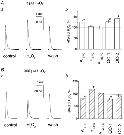
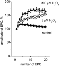
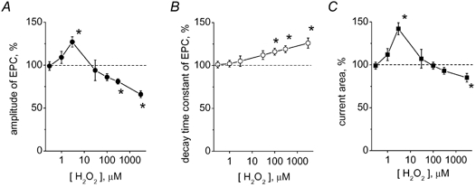
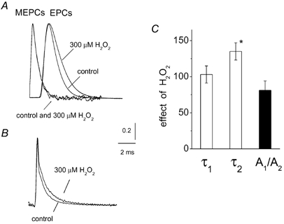
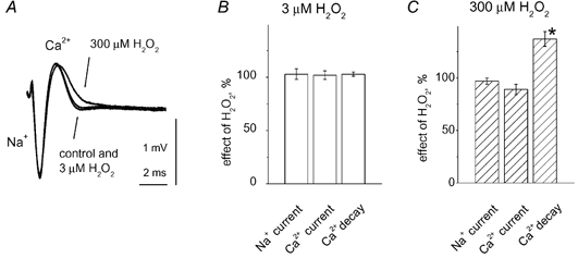
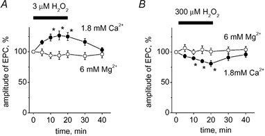
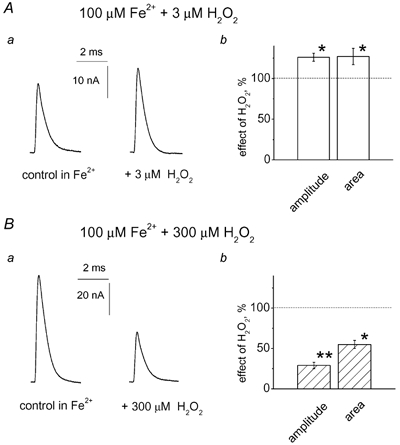
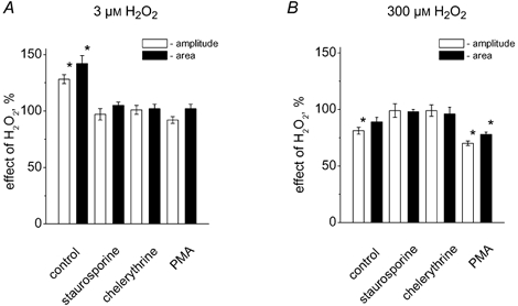
There is evidence that reactive oxygen species (ROS) are produced and released during neuromuscular activity, but their role in synaptic transmission is not known. Using a two-electrode voltage-clamp technique, at frog neuromuscular junctions, the action H2O2 on end-plate currents (EPC) was studied to determine the targets for this membrane-permeable ROS. In curarized or cut muscles, micromolar concentrations of H2O2 increased the amplitude of EPCs. Higher (> 30 microM) doses inhibited EPCs and prolonged current decay. These effects were presynaptic since H2O2 did not change the amplitude or duration of miniature EPCs (although it reduced the rate of spontaneous release at high concentrations). Quantal analysis and deconvolution methods showed that facilitation of EPCs was due to increased quantal release, while depression was accompanied by temporal dispersion of evoked release. Extracellular recordings revealed prolonged presynaptic Ca2+ entry in the presence of high H2O2. Both low and high H2O2 increased presynaptic potentiation during high-frequency stimulation. Pro-oxidant Fe2+ did not affect facilitation by low doses of H2O2 but augmented the inhibition of EPCs by high H2O2, indicating involvement of hydroxyl radicals. High Mg2+ and the ROS scavenger N-acetylcysteine eliminated both the facilitatory and depressant effects of H2O2. The facilitatory effect of H2O2 was prevented by protein kinase C (PKC) inhibitors and 4beta-phorbol 12-myristate, 13-acetate (PMA), an activator of PKC. PKC inhibitors but not PMA also abolished the depressant effect of H2O2. Our data suggest complex presynaptic actions of H2O2, which could serve as a fast feedback modulator of intense neuromuscular transmission.


Figures

Figure 1
Figure 1. Opposite effects of low and high concentrations of H2O2 on multiquantal end-plate currents (EPCs)
A, facilitating effect of 3 μM H2O2 on multiquantal EPCs. Aa, representative EPCs in control, in the presence of 3 μM H2O2 and after washout. Ab, histograms showing the action of 3 μM H2O2 on peak amplitude of EPCs (AEPC), decay time constant of EPCs (τEPC), amplitude of MEPCs (AMEPC) and quantal content estimated either as the ratio of peak current amplitudes of averaged EPCs and MEPCs (QC-1) or as the ratio of current integrals (QC-2) on cut preparation (* P < 0.05; n = 6). Data in this and subsequent histograms are presented as the mean ± s.e.m. (n = number of synapses). B, depressant action of 300 μM H2O2 on multiquantal EPCs. Ba, EPCs in control, in the presence of 300 μM H2O2 and after washout. Bb, histograms showing the action of 300 μM H2O2 on the same parameters of EPCs and MEPCs as in Ab (* P < 0.05; n = 6). Note that depression of QC-1 was stronger (P < 0.05) than that of QC-2 (P > 0.05).
Figure 3
Figure 3. The action of H2O2 on short-term plasticity
A, peak amplitudes of 20 successive EPCs during 60 Hz motor nerve stimulation of a cut preparation in control (•), and in the presence of 3 μM (○) or 300 μM (▴; n = 5-7) H2O2. Note the significantly enhanced potentiation of successive EPCs in the presence of both concentrations of H2O2.
Figure 2
Figure 2. Concentration dependence of H2O2 action on EPCs
A, effect of various H2O2 concentrations on peak EPC amplitude in a cut preparation. B, effect of H2O2 on the decay time constant of EPCs. C, the action of H2O2 on the current integrals. (* P < 0.05; n = 4-8 synapses). Note the biphasic action of H2O2 on the amplitude and area of EPCs and monophasic action of H2O2 on the time course of EPCs.
Figure 4
Figure 4. Effect of H2O2 on the time course of transmitter release
A, examples of averaged MEPCs (n = 120) or averaged multiquantal EPCs (n = 10) used for deconvolution in control and after 300 μM H2O2. All signals were normalized for comparison. Note that H2O2 selectively prolonged the decay of EPCs, while in the presence of 300 μM H2O2 the shape of MEPCs was the same as in control. B illustrates the results of deconvolution of the averaged EPC from A (transmitter release functions) with the fitted MEPCs (for detail see Methods). Deconvolution revealed two exponential components of transmitter release with decay time constants of 0.18 ± 0.04 and 1.02 ± 0.11 ms, respectively. Transmitter release functions were obtained by averaging data from 6 endplates. Note that 300 μM H2O2 increased the relative contribution of the slow phase of transmitter release function. C, histograms showing that 300 μM H2O2 significantly increased the decay time constant of the slow phase of the release function (τ2; * P < 0.05; n = 6), while it did not affect the fast phase (τ1; P > 0.05; n = 6) or the ratio of the amplitudes of the two components (A1/A2; P > 0.05; n = 6).
Figure 5
Figure 5. Effect of H2O2 on the presynaptic Ca2+ current
A, presynaptic action potential recorded by a microelectrode inserted into the perineurium adjacent to the nerve ending. The large initial downward part of the action potential represents the initial Na+ current while the late upward part represents the Ca2+ current. Each signal was obtained by averaging 24 records evoked at 0.05 Hz. The postsynaptic response was blocked by a high dose of (+)-tubocurarine (for details, see Methods). Low (3 μM) H2O2 did not change the Na+ or Ca2+ phase of the presynaptic signal, while high (100 μM) H2O2 slowed the decay of the Ca2+ current without producing significant changes in the Na+ phase. B and C, histograms showing the action of 3 or 300 μM H2O2 on the amplitude (% change vs. control) or the time constant of Ca2+ current decay. * P < 0.05, n = 6 for both H2O2 concentrations.
Figure 6
Figure 6. Modification by Mg2+ of H2O2 action on EPCs
A, time profile for the facilitatory effect of 3 μM H2O2 on EPCs in control (•; [Ca2+]o = 1.8 mM) and in a magnesium preparation (○; [Mg2+]o = 6 mM; [Ca2+]o = 0.9 mM). * P < 0.05; n = 5. B, time profile for the depressant effect of 300 μM H2O2 on EPCs in control (•) and in a magnesium preparation (○; * P < 0.05; n = 5).
Figure 7
Figure 7. Fe2+ selectively promoted the depressant effect of 300 μM H2O2
Aa, effect of 3 μM H2O2 in the presence of 100 μM Fe2+ on multiquantal EPCs in a cut preparation. Ab, histograms showing the facilitatory action of 3 μM H2O2 on peak EPC amplitude or current area. Note that the facilitatory action of a low dose of H2O2 was not changed by Fe2+ (26 ± 5 % enhancement in Fe2+; * P < 0.05; n = 5; compare with 28 ± 7 % in control). Ba, the depressant action of 300 μM H2O2 on EPCs was strongly enhanced by 100 μM Fe2+. Bb, histograms showing the action of 300 μM H2O2 on peak EPC amplitude or current area in the presence of 100 μM Fe2+ (** P < 0.01; significantly different from the action of 300 μM H2O2 in control; n = 5).
Figure 8
Figure 8. Testing the role of PKC in the action of H2O2
A, histograms showing the action of 3 μM H2O2 on the peak amplitude of EPCs (□) and the current area (▪) in control (* P < 0.05; n = 5), in the presence of 0.5 μM staurosporine (P > 0.05; n = 5), 10 μM chelerythrine (P > 0.05; n = 5), and 0.5 μM PMA (P > 0.05; n = 5). B, the same data for the action of 300 μM H2O2 (n = 4-7). Note, that unlike the facilitatory action of 3 μM H2O2, the depressant effect of 300 μM H2O2 was not only preserved in the presence of PMA but even enhanced compared with control (P < 0.05).

Similar articles

Cited by

References

    1. Auerbach JM, Segal M. Peroxide modulation of slow onset potentiation in rat hippocampus. J Neurosci. 1997;17:8695–8701. - PMC - PubMed
    1. Barstad JA, Lilleheil G. Transversaly cut diaphragm preparation from rat. An adjuvant tool in the study of the physiology and pharmacology of the myoneural junction. Arch Int Pharmacodyn Ther. 1968;175:373–390. - PubMed
    1. Boveris A, Oshino N, Chance B. The cellular production of hydrogen peroxide. Biochem J. 1972;128:617–630. - PMC - PubMed
    1. Bukcharaeva EA, Kim KC, Moravec J, Nikolsky EE, Vyskocil F. Noradrenaline synchronizes evoked quantal release at frog neuromuscular junctions. J Physiol. 1999;517:879–888. - PMC - PubMed
    1. Castonguay A, Lévesque S, Robitaille R. Glial cells as active partners in synaptic functions. Prog Brain Res. 2001;132:227–240. - PubMed

Publication types

MeSH terms

LinkOut - more resources