. 2003 Aug;112(3):379-88.
doi: 10.1172/JCI18038.

A mutant, noninhibitory plasminogen activator inhibitor type 1 decreases matrix accumulation in experimental glomerulonephritis

Affiliations

A mutant, noninhibitory plasminogen activator inhibitor type 1 decreases matrix accumulation in experimental glomerulonephritis

Yufeng Huang et al. J Clin Invest. 2003 Aug.

Abstract

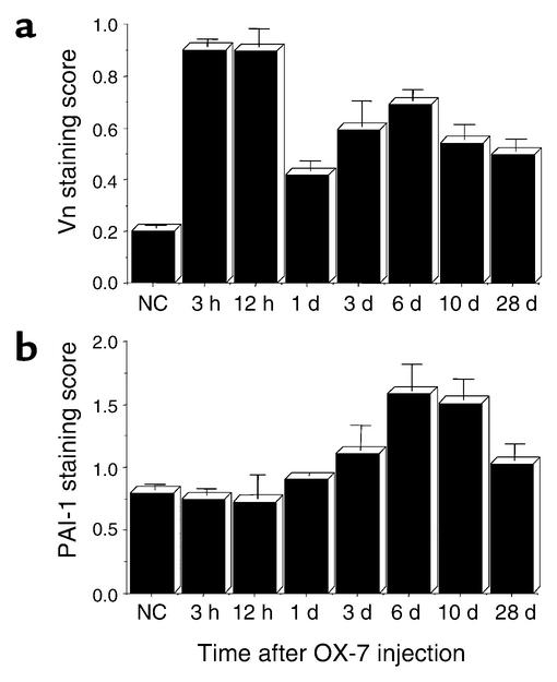
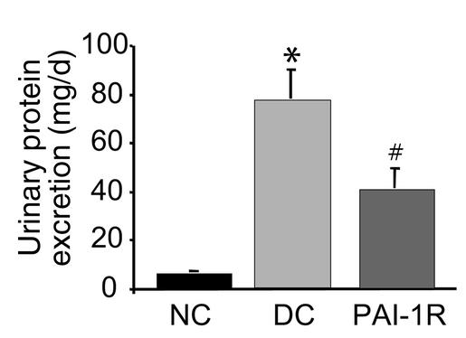
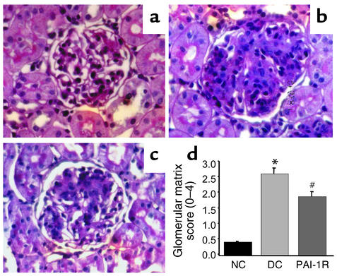
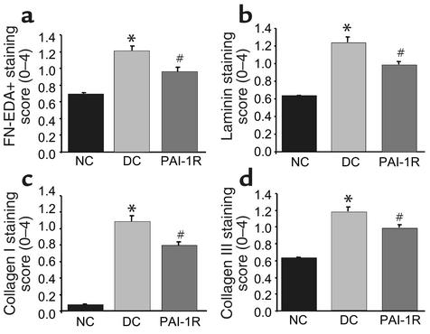
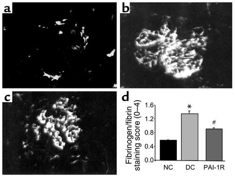
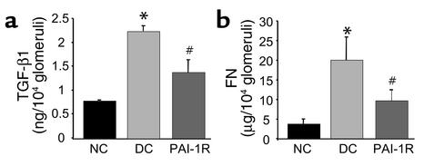
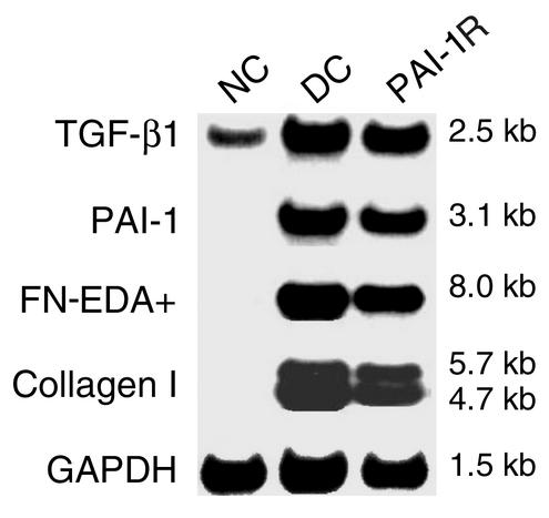
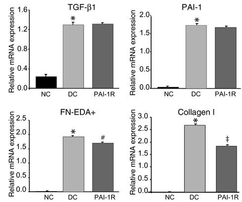
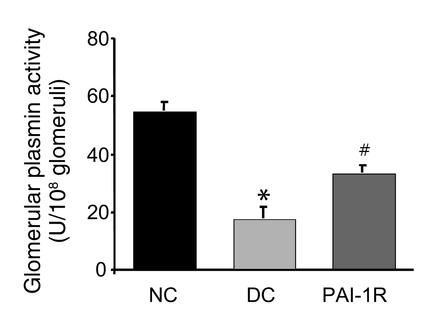
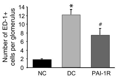
In fibrotic renal disease, elevated TGF-beta and angiotensin II lead to increased plasminogen activator inhibitor type 1 (PAI-1). PAI-1 appears to reduce glomerular mesangial matrix turnover by inhibiting plasminogen activators, thereby decreasing plasmin generation and plasmin-mediated matrix degradation. We hypothesized that therapy with a mutant human PAI-1 (PAI-1R) that binds to matrix vitronectin but does not inhibit plasminogen activators, would enhance plasmin generation, increase matrix turnover, and decrease matrix accumulation in experimental glomerulonephritis. Three experimental groups included normal, untreated disease control, and PAI-1R-treated nephritic rats. Plasmin generation by isolated day 3 glomeruli was dramatically decreased by 69%, a decrease that was reversed 43% (P < 0.02) by in vivo PAI-1R treatment. At day 6, animals treated with PAI-1R showed significant reductions in proteinuria (48%, P < 0.02), glomerular staining for periodic acid-Schiff positive material (33%, P < 0.02), collagen I (28%, P < 0.01), collagen III (34%, P < 0.01), fibronectin (48%, P < 0.01), and laminin (41%, P < 0.01), and in collagen I (P < 0.01) and fibronectin mRNA levels (P < 0.02). Treatment did not alter overexpression of TGF-beta1 and PAI-1 mRNAs, although TGF-beta1 protein was significantly reduced. These observations strongly support our hypothesis that PAI-1R reduces glomerulosclerosis by competing with endogenous PAI-1, restoring plasmin generation, inhibiting inflammatory cell infiltration, decreasing local TGF-beta1 concentration, and reducing matrix accumulation.


Figures

Figure 1
Figure 1
Time course of glomerular Vn (a) and endogenous PAI-1 (b) staining in OX-7–induced nephritis. Data are from three rats at each timepoint. NC, normal control.
Figure 2
Figure 2
Time course of disappearance of PAI-1R from nephritic glomeruli, shown by injected PAI-1R staining in OX-7–induced nephritic glomeruli at d1. Representative photomicrograph of glomeruli from two rats at each timepoint injected with PAI-1R at 1 mg/kg body weight.
Figure 3
Figure 3
Colocalization of PAI-1R with Vn. A glomerulus from a d1 animal sacrificed 1 hour after PAI-1R injection. Staining for (a) rat Vn (red) and (b) PAI-1R (green). (c) Double staining for Vn and PAI-1R. Injected PAI-1R colocalized with endogenous rat Vn in the mesangium. Normal control (d) and disease control (e) rats showed Vn staining only in glomerulus, and no staining for human PAI-1 in kidney.
Figure 4
Figure 4
Effects of PAI-1R on 24-hour urinary protein excretion from d5 to d6. Urinary protein excretion was significantly lower in the PAI-1R–treated, nephritic group. *P < 0.001 vs. normal control. #P < 0.02 vs. disease control (DC).
Figure 5
Figure 5
Glomerular histology. Representative photomicrographs of glomeruli from normal control rats (a), disease control rats treated with PBS (b), and PAI-1R–treated, nephritic rats (c) at d6. (d) Graphic representation of PAS staining scores of each group. PAI-1R treatment resulted in a significant reduction in PAS staining score compared with disease control rats. *P < 0.001 vs. normal control; #P < 0.02 vs. disease control.
Figure 6
Figure 6
Immunofluorescent staining score for ECM proteins at d6. Glomerular staining for FN-EDA+ (a), laminin (b), type I collagen (c), and type III collagen (d) were lower in the PAI-1R–treated, nephritic group. *P < 0.001 compared with normal control. #P < 0.01 compared with disease control.
Figure 7
Figure 7
Immunofluorescent staining for glomerular fibrinogen/fibrin at d6. Representative photomicrograph of a normal rat (a), a disease control (b), and a PAI-1R–treated, nephritic rat (c). Graphic representation of fibrinogen/fibrin staining scores for each group (d). PAI-1R treatment resulted in a significant reduction in glomerular fibrin deposition compared with disease control rats. *P < 0.001 vs. normal control; #P < 0.001 vs. disease control.
Figure 8
Figure 8
Effects of PAI-1R on TGF-β1 and fibronectin (FN) content in glomeruli at d6. The glomerular content of total TGF-β1 (a) and fibronectin (b) was significantly lower in the PAI-1R–treated group. *P < 0.001 vs. normal control; #P < 0.01 vs. disease control.
Figure 9
Figure 9
Effects of PAI-1R treatment on glomerular mRNA expression in anti–thy-1 nephritis at d6. Representative Northern blot is shown.
Figure 10
Figure 10
Relative glomerular mRNA expression of TGF-β1, PAI-1, fibronectin, and type I collagen. Glomerular mRNA levels of fibronectin and type I collagen were significantly reduced at d6, but the overexpression of TGF-β1 and PAI-1 was not changed in the PAI-1R–treated group. *P < 0.001 vs. normal control; #P < 0.02 vs. disease control; P < 0.01 vs. disease control.
Figure 11
Figure 11
Effects of PAI-1R on the glomerular plasmin activity of nephritic rats at d3. Kidneys were removed 10 minutes after PBS or PAI-1R injection (1 mg/kg body weight). Nephritic glomeruli had decreased plasmin activity, which was elevated by in vivo presacrifice injection of PAI-1R (n = 4 rats per group). *P < 0.01 vs. normal control; #P < 0.02 vs. disease control.
Figure 12
Figure 12
Number of monocytes/macrophages infiltrating glomeruli in anti–thy-1 nephritis at d6. PAI-1R treatment resulted in a significant reduction in monocyte/macrophage infiltration compared with disease control rats. *P < 0.001 vs. normal control; #P < 0.02 vs. disease control.

Comment in

References

    1. Border WA, Noble NA. Transforming growth factor beta in tissue fibrosis. N. Engl. J. Med. 1994;331:1286–1292. - PubMed
    1. Border WA, Okuda S, Nakamura T. Extracellular matrix and glomerular disease. Semin. Nephrol. 1989;9:307–317. - PubMed
    1. Border WA, Noble NA. Interactions of transforming growth factor-beta and angiotensin II in renal fibrosis. Hypertension. 1998;31:181–188. - PubMed
    1. Schnaper HW. Balance between matrix synthesis and degradation: a determinant of glomerulosclerosis. Pediatr. Nephrol. 1995;9:104–111. - PubMed
    1. Gaedeke J, Peters H, Noble NA, Border WA. Angiotensin II, TGF-β and renal fibrosis. Contrib. Nephrol. 2001;135:153–160. - PubMed

Publication types

MeSH terms