. 2003 Aug;112(3):450-6.
doi: 10.1172/JCI18264.

Griscelli syndrome restricted to hypopigmentation results from a melanophilin defect (GS3) or a MYO5A F-exon deletion (GS1)

Affiliations

Griscelli syndrome restricted to hypopigmentation results from a melanophilin defect (GS3) or a MYO5A F-exon deletion (GS1)

Gaël Ménasché et al. J Clin Invest. 2003 Aug.

Erratum in

  • J Clin Invest. 2005 Apr;115(4):1100

Abstract

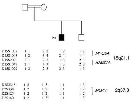
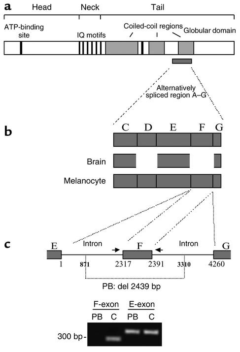
Griscelli syndrome (GS) is a rare autosomal recessive disorder that associates hypopigmentation, characterized by a silver-gray sheen of the hair and the presence of large clusters of pigment in the hair shaft, and the occurrence of either a primary neurological impairment or a severe immune disorder. Two different genetic forms, GS1 and GS2, respectively, account for the mutually exclusive neurological and immunological phenotypes. Mutations in the gene encoding the molecular motor protein Myosin Va (MyoVa) cause GS1 and the dilute mutant in mice, whereas mutations in the gene encoding the small GTPase Rab27a are responsible for GS2 and the ashen mouse model. We herein present genetic and functional evidence that a third form of GS (GS3), whose expression is restricted to the characteristic hypopigmentation of GS, results from mutation in the gene that encodes melanophilin (Mlph), the ortholog of the gene mutated in leaden mice. We also show that an identical phenotype can result from the deletion of the MYO5A F-exon, an exon with a tissue-restricted expression pattern. This spectrum of GS conditions pinpoints the distinct molecular pathways used by melanocytes, neurons, and immune cells in secretory granule exocytosis, which in part remain to be unraveled.


Figures

Figure 1
Figure 1
Light microscopy of patients’ hair shafts. Typical features of GS are the large clumps of pigment irregularly distributed along the hair shaft, as shown for a GS1 patient and a GS2 patient. The same aspect is observed in the hair shafts of PA and PB. In contrast, a fine, evenly distributed pigment is observed in the control hair shaft.
Figure 2
Figure 2
Linkage analysis in PA’s family tree. PA’s family tree and haplotype analysis of polymorphic markers spanning the GS locus (with MYO5A and RAB27A) on chromosome 15q21.1, and the MLPH locus on chromosome 2q37.3. Haplotype segregation in PA’s family shows an absence of linkage of the GS to the MYO5A/RAB27A locus, whereas a homozygous haplotype segregates in the patient at the MLPH locus.
Figure 3
Figure 3
MLPH missense mutation in PA. (a) Detection of MLPH mutation in PA’s family by fluorometric sequencing. DNA sequence analysis of exon 1 shows a C-to-T transition resulting in an R35W substitution in PA. The mutations of the mother and father are heterozygous for R35W. (b) Sequence alignment of the SHD1 domain of human Mlph (MLPH) and mouse Mlph (Mlph) as well as of mouse Rabphilin-3A (mRabphilin-3A). The residue R35 substituted by W in PA is indicated by an arrow, and the seven residues deleted in the leaden mutant are indicated by italics highlighted in gray. The Rabphilin-3A residues mediating the contact with Rab3A (26) are indicated by asterisks. Amino acid numbers are indicated at the right end of each line.
Figure 4
Figure 4
Association of Mlph mutant proteins with Rab27a. (a) MLPH mutation in PA affects Mlph-Rab27a interaction, as shown by coimmunoprecipitation analysis. WT Mlph (the MLPH sequence encoding the first 146 amino acids [SHD], cloned in pcDNA 3.1/Myc vector) or the mutant Mlph R35W, and Rab27a (the entire sequence encoding Rab27a, cloned in pFlag-CMV-4 vector), were cotransfected into 293T cells, immunoprecipitated with an anti-Flag antibody, and revealed with an anti-Myc antibody (IP: anti-Flag; Blot: anti-Myc). A similar amount of proteins was present (top and bottom panels). The positions of the marker (× 10–3) are shown on the left side. (b) None of the new substitutions introduced at position 35 of Mlph could restore Mlph-Rab27a interaction. The R35K, R35V, and R35F mutants were introduced in the SHD of Mlph by directed mutagenesis. Coimmunoprecipitated Flag-Rab27a detected by the anti-Flag antibody (IP: anti-Myc; Blot: anti-Flag) was only observed with the WT Mlph. The same blot was then stripped and reprobed with an anti-Myc antibody. The top panel indicates the total amount of expressed Flag-Rab27a (1:100 volume of the reaction mixture) used for immunoprecipitation. None of the mutant constructs was able to restore binding to Rab27a.
Figure 5
Figure 5
Overexpression of WT-SHD Mlph but not R35W-SHD Mlph interferes with melanosome transport. Melan-a cells were transfected with plasmids encoding pEGFP-C2, which allows a soluble GFP expression (a), or with GFP-WT-SHD-Mlph (c) or GFP-R35W-SHD-Mlph (e). (b, d, and f) Images of transmitted light showing the melanosome distribution. Bars: 20 μm.
Figure 6
Figure 6
Characterization of the MYO5A mutation in PB. (a) Schematic representation of MyoVa with the details of the various domains as previously reported (9) and of the alternatively spliced A–G region. (b) Schematic representation of the alternative splice forms expressed in melanocyte and brain tissues. (c) Deletion 871–3310 in PB’s MYO5A gene disrupts the F-exon as well as its 5′ and 3′ flanking intronic sequences. Number 1 of the base pair corresponds to the first base of the 5′ F-intron sequence. PCR amplification of the E- and F-exons in PB and control (C) is shown. Primer pairs used (indicated by arrows for the F-exon) were previously described (9).
Figure 7
Figure 7
Scheme of the heterotrimeric protein complex involved in human melanosome transport. A defect in any of the proteins, MyoVa, Rab27a, or Mlph, leads to identical pigmentary dilution, found in the three forms of GS. The F-exon of MyoVa is required for MyoVa-Mlph interaction and the SHD of Mlph for Mlph-Rab27a interaction. The locations of the genetic defects identified in PA and PB are shown.

Similar articles

Cited by

References

    1. Griscelli C, et al. A syndrome associating partial albinism and immunodeficiency. Am. J. Med. 1978;65:691–702. - PubMed
    1. Pastural E, et al. Griscelli disease maps to chromosome 15q21 and is associated with mutations in the myosin-Va gene. Nat. Genet. 1997;16:289–292. - PubMed
    1. Ménasché G, et al. Mutations in RAB27A cause Griscelli syndrome associated with hemophagocytic syndrome. Nat. Genet. 2000;25:173–176. - PubMed
    1. Bahadoran P, et al. Rab27a. A key to melanosome transport in human melanocytes. J. Cell Biol. 2001;152:843–850. - PMC - PubMed
    1. Langford GM, Molyneaux BJ. Myosin V in the brain: mutations lead to neurological defects. Brain Res. Brain Res. Rev. 1998;28:1–8. - PubMed

Publication types

MeSH terms