. 2003 Aug;15(8):1717-27.
doi: 10.1105/tpc.012856.

The interaction of two homeobox genes, BREVIPEDICELLUS and PENNYWISE, regulates internode patterning in the Arabidopsis inflorescence

Affiliations

The interaction of two homeobox genes, BREVIPEDICELLUS and PENNYWISE, regulates internode patterning in the Arabidopsis inflorescence

Harley M S Smith et al. Plant Cell. 2003 Aug.

Abstract

Plant architecture results from the activity of the shoot apical meristem, which initiates leaves, internodes, and axillary meristems. KNOTTED1-like homeobox (KNOX) genes are expressed in specific patterns in the shoot apical meristem and play important roles in plant architecture. KNOX proteins interact with BEL1-like (BELL) homeodomain proteins and together bind a target sequence with high affinity. We have obtained a mutation in one of the Arabidopsis BELL genes, PENNYWISE (PNY), that appears phenotypically similar to the KNOX mutant brevipedicellus (bp). Both bp and pny have randomly shorter internodes and display a slight increase in the number of axillary branches. The double mutant shows a synergistic phenotype of extremely short internodes interspersed with long internodes and increased branching. PNY is expressed in inflorescence and floral meristems and overlaps with BP in a discrete domain of the inflorescence meristem where we propose the internode is patterned. The physical association of the PNY and BP proteins suggests that they participate in a complex that regulates early patterning events in the inflorescence meristem.


Figures

Figure 1.
Figure 1.
Characterization of the Loss-of-Function pny Alleles. (A) The amino acid sequence of PNY showing the MEINOX-interacting domain (underlined) and the three–amino acid loop extension homeodomain (boldface). (B) The homeodomain of PNY was aligned with three other BELL proteins, BEL1, ATH1, and KNOTTED INTERACTION PROTEIN (KIP). (C) Scheme of the PNY gene. White boxes represent exons, and black lines represent introns. Triangles depict the approximate T-DNA insertion sites. (D) RT-PCR analysis using PNY gene-specific primers. Amplification occurred only from wild-type tissue (lane 1) and not from the pny-40126 (lane 2) or pny-57747 (lane 3) tissue. Hybridization of a 32P-labeled PNY probe occurred in the wild-type sample only after PCR products were transferred to a nylon membrane (Blot-PNY). Primers to ACT8 show that approximately equal amounts were amplified.
Figure 2.
Figure 2.
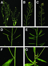
Characterization of the pny Phenotype. (A) Wild-type (left) and pny (right) plants showing that pny plants were slightly shorter. (B) Wild-type inflorescence with a regular pattern of internode development. (C) pny inflorescence with short and long internodes. (D) Close-up of a wild-type inflorescence. (E) Close-up of a pny inflorescence showing a cluster of siliques. (F) Paraclade in the axil of a cauline leaf on a wild-type plant. (G) Paraclade in the axil of a cauline leaf on a pny plant. The paraclades produced cauline leaves with no internode elongation between them (arrowheads).
Figure 3.
Figure 3.
Internode and Axillary Branch Defects. Measurements were taken from 10 plants from each genotype of the F2 progeny that resulted from a cross between pny-40126 and bp-9. (A) The average height of the wild type (Wt), pny, bp, and pny bp was determined, and the standard deviation was calculated. (B) The average number of paraclades that grew >20 mm was measured, and the standard deviation was determined. (C) Internode lengths between the first three paraclades (counting basipetally) were measured and grouped into 5-mm intervals. Wild type, dark blue; pny, red; bp, yellow; bp, pale blue. (D) Ten internodes between the 1st and 11th siliques (counting acropetally) were measured and graphed. Color code same as in (C).
Figure 4.
Figure 4.
Phenotype of the pny bp Double Mutant. (A) The inflorescence of the pny bp double mutant was very compact compared with those of the wild type (WT) and the bp and pny single mutants. (B) Close-up of the inflorescence of pny bp. (C) Paraclades from a pny bp plant were removed for photography. Clusters of siliques were visible on the main axis (m) and the paraclades (p). (D) The short internodes between the paraclades and the siliques in pny bp produced an aerial rosette. (E) Close-up view of a wild-type inflorescence. (F) Close-up view of a pny inflorescence. Note the clusters of siliques. (G) Close-up view of a bp inflorescence. Note the downward slant of the pedicels and the short internodes. (H) Close-up view of a pny bp inflorescence. Note the exaggerated clustering of siliques and short pedicels.
Figure 5.
Figure 5.
Histological Analysis of the Stems. (A) Cross-section through a wild-type stem. (B) Cross-section through a bp stem. (C) Cross section through a pny stem. (D) Cross-section through a pny bp stem. A band of phloem (blue-stained cells) formed beneath the cortex layer. (E) Close-up of a wild-type stem section. The vascular bundle, consisting of xylem and phloem, was detected inside of the cortex, protruding into the highly vacuolated pith cells. Cambial cells (opaque staining) were located between the xylem and phloem cells. (F) Close-up of a bp stem section. Bundles were closer together as a result of the overall reduction in size. The bundle indicated with an arrow contained small densely stained cells, suggesting that it was not fully differentiated. (G) Close-up of a pny stem section. Note the increase in the number of vascular bundles, as depicted by the blue patches of phloem. In addition, there was an increase in lignification (green-stained cells). (H) Close-up of a pny bp stem section. Note the thick continuous band of phloem cells. The cambium-like cells (opaque cells inside of the phloem) had random cell division patterns. Xylem elements were small and not lignified. Cortex and epidermal cells were larger, whereas pith cells were smaller. c, cortex; e, epidermis; i, interfascicular cells; ph, phloem; pi, pith; x, xylem. Bars = 1 mm in (A) to (D) and 500 nm in (E) to (H).
Figure 6.
Figure 6.
Expression Profile of PNY. RT-PCR was performed on different plant tissues (RT-PNY), which then were blotted to nylon and probed with a 32P-labeled PNY probe (Blot-PNY). PNY was amplified from inflorescence meristem tissue (M) that was obtained from ap1 cal double mutants. Only minute amounts of PNY could be amplified from seedlings (S) and mature leaves (L). No PCR product was detected in mature flowers (F), siliques (Si), stems and pedicles (SP), or roots (R). Approximately equal amounts of ACT8 PCR product (RT-ACT) were amplified from the tissues examined.
Figure 7.
Figure 7.
In Situ Hybridization of PNY and BP in Wild-Type Inflorescence Tissues. (A) and (B) Longitudinal cross-sections through the inflorescence meristem. PNY mRNA was localized in stripes of cells on the flanks of the meristem (arrows). (C) and (D) Transverse sections through the inflorescence meristem. PNY localized to bands of cells that may encircle initiating floral primordia. Note that PNY was not expressed in early floral primordia but was expressed at a later stage of flower development. (E) and (F) BP was expressed in a band of cells on the flanks of the meristem (arrows). In addition, BP was expressed at the base of the floral primordia four to five cells from the epidermal layer. BP also localized to cortex cells adjacent to vascular tissue in elongating internodes and pedicels. (G) and (H) PNY mRNA localized to cells in the floral meristem but not in developing floral organ primordia (arrows). c, cortex; fm, floral meristem; im, inflorescence meristem; pd, pedicle. Bars = 100 nm.
Figure 8.
Figure 8.
Ligand Blot Analysis of PNY. (A) PNY was labeled in vitro with 35S-Met, separated by SDS-PAGE, and analyzed by autoradiography. The molecular mass of PNY is 67 kD (arrowhead). (B) The MEINOX (M) domain of STM (lane 2), BP (lane 3), and KNAT2 (lane 4) was fused to GST, and ∼1 μg of full-length recombinant expressed and purified proteins was separated by SDS-PAGE. The molecular mass of the full-length GST fusions was 61 kD for GST-MSTM, 59 kD for GST-MBP, and 41 kD for GST-MKNAT2. Other protein products detected in each lane correspond to partially degraded fusion proteins or bacterial proteins that copurify with the GST fusion proteins. These protein products also serve as internal negative controls, because the interaction of KNOX and BELL proteins requires an intact MEINOX domain (our unpublished data). GST and BSA were used as negative controls. (C) Ligand blots probed with radiolabeled PNY and analyzed by autoradiography demonstrated that PNY interacted with GST-MSTM and GST-MBP, whereas little or no binding was detected for GST-MKNAT2, GST, or BSA.

Similar articles

Cited by

References

    1. Bellaoui, M., Pidkowich, M.S., Samach, A., Kushalappa, K., Kohalmi, S.E., Modrusan, Z., Crosby, W.L., and Haughn, G.W. (2001). The Arabidopsis BELL1 and KNOX TALE homeodomain proteins interact through a domain conserved between plants and animals. Plant Cell 11, 2455–2470. - PMC - PubMed
    1. Chen, M.H., Sheng, J., Hind, G., Handa, A.K., and Citovsky, V. (2000). Interaction between the tobacco mosaic virus movement protein and host cell pectin methylesterases is required for viral cell-to-cell movement. EMBO J. 19, 913–920. - PMC - PubMed
    1. Cline, M.G. (1994). The role of hormones in apical dominance: New approaches to an old problem in plant development. Physiol. Plant. 90, 230–237.
    1. Cutter, E.G. (1971). Plant Anatomy: Experiment and Interpretation. (London: Edward Arnold).
    1. Digby, J., and Wareing, P.F. (1966). The effect of applied growth hormones on cambial activity and the differentiation of the cambial derivatives. Ann. Bot. 30, 539–548.

Publication types

Associated data