Comparative Study
. 2003 Aug;15(8):1728-39.
doi: 10.1105/tpc.012401.

The MSP1 gene is necessary to restrict the number of cells entering into male and female sporogenesis and to initiate anther wall formation in rice

Affiliations
Comparative Study

The MSP1 gene is necessary to restrict the number of cells entering into male and female sporogenesis and to initiate anther wall formation in rice

Ken-Ichi Nonomura et al. Plant Cell. 2003 Aug.

Abstract

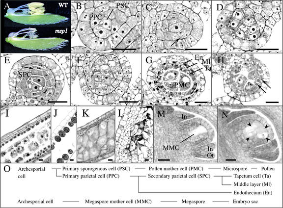
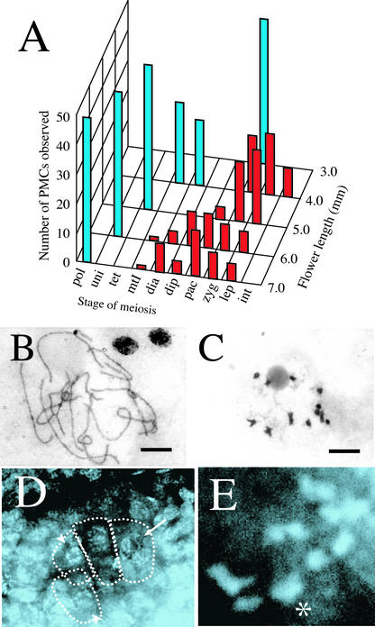
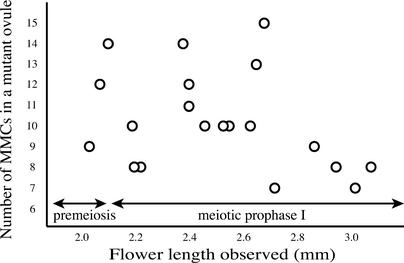
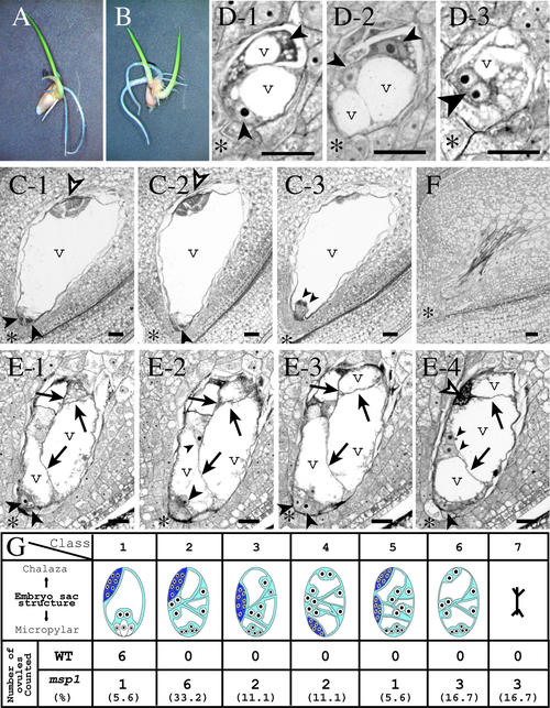
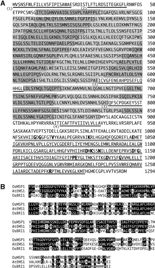
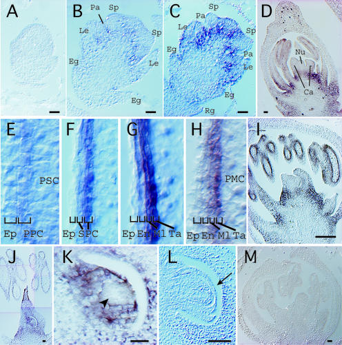
The function of the novel gene MSP1 (MULTIPLE SPOROCYTE), which controls early sporogenic development, was elucidated by characterizing a retrotransposon-tagged mutation of rice. The MSP1 gene encoded a Leu-rich repeat receptor-like protein kinase. The msp1 mutation gave rise to an excessive number of both male and female sporocytes. In addition, the formation of anther wall layers was disordered and the tapetum layer was lost completely. Although the mutation never affected homologous chromosome pairing and chiasma maintenance, the development of pollen mother cells was arrested at various stages of meiotic prophase I, which resulted in complete male sterility. Meanwhile, plural megaspore mother cells in a mutant ovule generated several megaspores, underwent gametogenesis, and produced germinable seeds when fertilized with wild-type pollen despite disorganized female gametophytes. In situ expression of MSP1 was detected in surrounding cells of male and female sporocytes and some flower tissues, but never in the sporocytes themselves. These results suggest that the MSP1 product plays crucial roles in restricting the number of cells entering into male and female sporogenesis and in initiating anther wall formation in rice.


Figures

Figure 1.
Figure 1.
Histological Features of Male and Female Reproductive Organ Development in the Wild Type and the msp1 Mutant. (A) Flower and anther morphology of the wild type (WT) and msp1 at the heading stage. (B) to (H) Transverse sections of the anther. (B) PSCs and PPCs were already differentiated in a wild-type anther at stage II. (C) PPCs began to divide periclinally (arrow) to differentiate secondary parietal cells in a wild-type anther at stage IV. (D) Cell mass was increased in the mutant microsporangia, but no layered structure was observed at stage IV. (E) Secondary parietal cells (SPC) began to divide periclinally (arrow) in a wild-type anther at stage V. (F) Cells of a similar size occupied the mutant microsporangia at stage V. (G) PMCs, tapetum cells (Ta), the middle layer (Ml), and the endothecium (En) were established in a wild-type anther in meiotic prophase I. (H) A layered structure, but with irregular proliferation, appeared in a mutant anther in meiotic prophase I (arrows). (I) to (N) Longitudinal sections of the anther and the ovule. (I) A densely stained tapetum layer located at the fourth inner layer was observed clearly in a longitudinal section of a wild-type anther in meiotic prophase I. (J) Upon the completion of pollen grain maturation, inner wall layers were degraded in a wild-type anther. (K) Tapetum cells were never observed in the longitudinal section of a mutant anther in meiotic prophase I. (L) Cells of a similar size formed a layer-like structure. PMCs were degraded in the mutant at the stage corresponding to the pollen maturation in the wild type shown in (J). (M) A single and elongated MMC was observed in a wild-type nucellus with developing inner integuments (In) and outer integuments (Ot). (N) At least four MMCs (arrowheads) were observed in a mutant nucellus with normally developing inner and outer integuments. (O) Wild-type cell lineage of male and female germ cells. Bars = 20 μm.
Figure 3.
Figure 3.
msp1 PMCs and MMCs Underwent Normal Synapsis and Chiasma Maintenance in Meiosis. (A) Histogram showing the number of PMCs in each meiotic stage investigated in flowers from 3.0 to 7.0 mm long. Blue and red bars indicate the number of PMCs in wild-type and mutant anthers, respectively. Stages of meiosis are from right to left: int, premeiotic interphase; lep, leptotene; zyg, zygotene; pac, pachytene; dip, diplotene; dia, diakinesis; mtI, metaphase I; uni, uninucleate; pol, matured pollen. (B) A complete set of 12 homologous chromosome pairs observed in the mutant PMC in pachytene. Bar = 10 μm. (C) Twelve bivalent chromosomes maintaining normal chiasmata in the mutant PMC in diakinesis. Bar = 10 μm. (D) An optical and longitudinal section of the mutant ovule. Several meiotic nuclei in pachytene (arrow) and diakinesis (arrowhead) were observed in the nucellus. (E) Magnified view of an MMC. A complete set of 12 homologous pairs in diakinesis was observed. The asterisk indicates the two overlapping pairs.
Figure 2.
Figure 2.
Relationship between the Number of MMCs in a msp1 Ovule and the Progression of MMC Development. The number of MMCs and their cell stages were observed using a series of longitudinal sections derived from a mutant ovule. From the very early stages of ovule development, the mutant nucellus contained multiple MMCs, suggesting that a discrete group of plural precursor cells was differentiated into archesporial cells in a mutant ovule, rather than a single archesporial cell proliferated abnormally to produce supernumerary MMCs. The correlation of meiosis progression to flower elongation was reported by Miyoshi et al. (2001).
Figure 4.
Figure 4.
Aberrant Megagametogenesis in the msp1 Ovule. (A) A germinated hybrid seed with a single embryo derived from the msp1 mutant crossed with wild-type pollen. (B) A germinated hybrid seed with twin embryos. (C) to (E) Longitudinal sections of wild-type and mutant ovules. (C) A series of three sections from a wild-type ovule. The egg apparatus cells (an egg cell and two synergids) appeared at the micropylar end (large arrowheads), polar nuclei were close to the egg apparatus (small arrowheads), and an antipodal tissue was at the chalazal end (open arrowheads). A central cell is mostly occupied by a vacuole (v). Asterisks indicate the micropylar end. (D) A series of three sections from a single mutant ovule containing multiple megaspores. At least four megaspores were included in the ovule (small and large arrowheads). The binucleated spore (large arrowhead) suggested that the spore had just entered into the first mitotic division of megagametogenesis. (E) A series of four sections from a mutant ovule. This ovule generated a single embryo sac but was compartmented by cell walls (arrows). Various numbers of cells were observed at the micropylar end (arrowheads). In this ovule, an antipodal tissue (open arrowheads) developed at a normal position. (F) The mutant ovule, empty and collapsed. (G) Embryo sac composition of the wild type (WT) and the mutant. The embryo sac morphology was classified into seven groups (from left to right): class 1 exhibited a normal configuration; classes 2 to 6 developed an embryo sac with aberrantly internal walls and various numbers of nuclei, but the position of the antipodal tissue developed differently as follows: class 2, at the normal position; class 3, at the middle portion; class 4, at the micropylar end; class 5, duplicated at the chalazal and middle portions; class 6, no antipodal tissue was developed. In class 7, the ovule was empty and collapsed. The frequency is presented at the bottom of each classification. Bars = 20 μm.
Figure 5.
Figure 5.
Identification and Characterization of the MSP1 Gene. (A) DNA gel blot analysis of some of 53 progeny of the msp1 heterozygotes. A Tos17 insertion induced a 4.0-kb polymorphic band, whereas the wild type showed a 4.3-kb band (lane P). The 4.0-kb bands were linked completely with the sterile phenotype (S). Homozygosity (W) or heterozygosity (H) of fertile plants was determined in the next generation. (B) Genomic structure of the MSP1 gene. The sequences corresponding to the cDNA are shown as closed boxes. Vertical arrows indicate the Tos17 insert site found in three alleles. ATG and TAA indicate start and stop codons, respectively. A pair of horizontal arrows and three lines below the gene structure indicate the positions of primers for RT-PCR and probes for DNA gel blot analysis and the in situ hybridization experiment, respectively. Kp, KpnI; Xb, XbaI; Xh, XhoI. (C) DNA gel blot analysis of cv Nipponbare genomic DNA using a probe shown in (B). (D) Flowers of complemented msp1 with the 7.7-kb XbaI-BamHI fragment containing the entire MSP1 gene. The two flowers are from different transformations. (E) An msp1 flower transformed with only a vector sequence having sterile anthers. (F) MSP1 expression analyzed by RT-PCR with (+) and without (−) reverse transcriptase. Amplified fragments were provided for ethidium bromide staining (top gel) and for DNA gel blot analysis (middle gel). RT-PCR for a rice actin gene (RAc) is shown as a positive control (bottom gel). Lane C, a positive control using cDNA as a PCR template.
Figure 6.
Figure 6.
The MSP1 Gene Encodes an LRR Receptor–Like Protein Kinase. (A) The shaded bars (light and dark shading is used alternately) indicate 34 LRR units. The underlined sequences indicate (1) a predicted signal peptide, (2) a putative Leu zipper motif, (3) the islands of 21 and 13 amino acids separating LRRs, and (4) a putative transmembrane domain. The double underline indicates a putative kinase domain. Boldface letters indicate invariant amino acid residues conserved completely in all Ser/Thr kinases (Hanks and Quinn, 1991). (B) Alignment of the kinase domains of OsMSP1, AtEMS1, and OsBRI1. Sequences were aligned with the CLUSTAL W program (http://www.ddbj.nig.ac.jp). Residues identical to those of OsMSP1 are highlighted in black.
Figure 7.
Figure 7.
In Situ Expression of the MSP1 Gene in Reproductive Organs of a Wild-Type Flower. (A) A palea-primordium differentiation stage. (B) An early differentiation stage of stamen and pistil primordia. The signal began to appear at the basal regions of each organ. Eg, empty glume; Le, lemma; Pa, palea; Sp, stamen primordium. (C) A late differentiation stage of stamen and pistil primordia. Rg, rudimentary glume. (D) A young flower at stage II. MSP1 signals began to appear in the inner wall layer of anthers, but no signal was observed in the female tissue. Ca, carpel; Nu, nucellus. (E) to (H) Magnified views of the inner walls formed in developing anthers from earlier (E) to later (H) stages. The strongest MSP1 expression was detected in the innermost cell layer attached to the sporogenous cells or sporocytes. Ep, epidermis; En, endothecium; Ml, middle layer; SPC, secondary parietal cells; Ta, tapetum cells. (I) A young flower at the stage of PMCs entering into meiotic prophase. No MSP1 signal was observed in the female tissue. (J) MSP1 expression was observed in a whole pistil, whereas expression in the anther had already disappeared. (K) A magnified view of the nucellus in (J). The nucellar cells were stained except for the MMC region indicated by the arrowhead. (L) MSP1 expression disappeared in the nucellus wrapped by the inner integuments (arrow). (M) Control experiment at the stage corresponding to (I) using a sense probe. Bars = 20 μm.

Similar articles

Cited by

References

    1. Bhojwani, S.S., and Bhatnagar, S.P. (1992). The Embryology of Angiosperms. (New Delhi: Vikas Publishing House).
    1. Canales, C., Bhatt, A.M., Scott, R., and Dickinson, H. (2002). EXS, a putative LRR receptor kinase, regulates male germline cell number and tapetal identity and promotes seed development in Arabidopsis. Curr. Biol. 12, 1718–1727. - PubMed
    1. Chapman, G.P. (1987). The tapetum. Int. Rev. Cytol. 107, 111–125.
    1. Chaubal, R., Zanella, C., Trimnell, M.R., Fox, T.W., Albertsen, M.C., and Bedinger, P. (2000). Two male-sterile mutants of Zea mays (Poaceae) with an extra cell division in the anther wall. Am. J. Bot. 87, 1193–1201. - PubMed
    1. Clark, S.E. (2001). Cell signalling at the shoot meristem. Nat. Rev. Mol. Cell Biol. 2, 276–284. - PubMed

Publication types

Substances

Associated data

LinkOut - more resources