. 2003 Aug;15(8):1740-8.
doi: 10.1105/tpc.012815.

Control of cellulose synthase complex localization in developing xylem

Affiliations

Control of cellulose synthase complex localization in developing xylem

John C Gardiner et al. Plant Cell. 2003 Aug.

Abstract

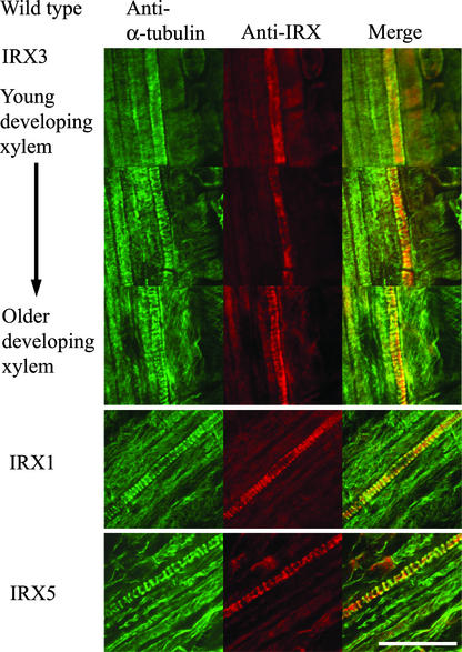
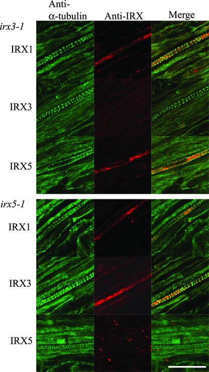
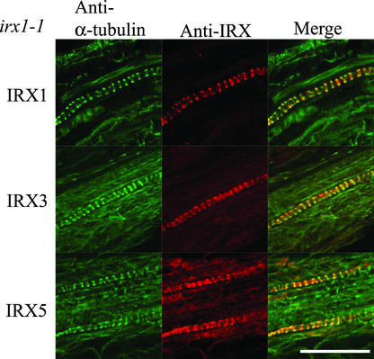
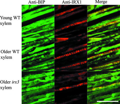
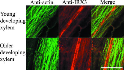
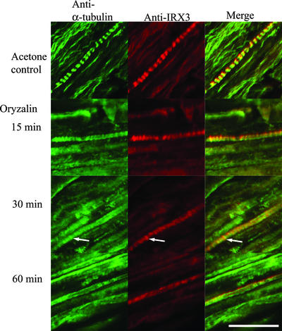
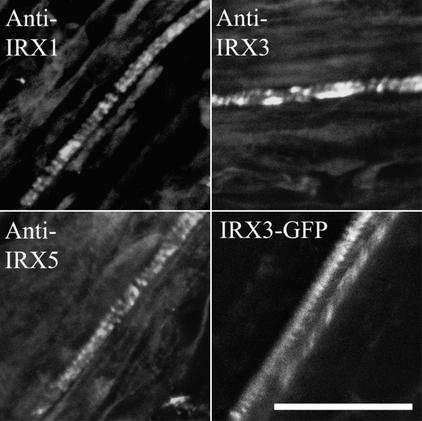
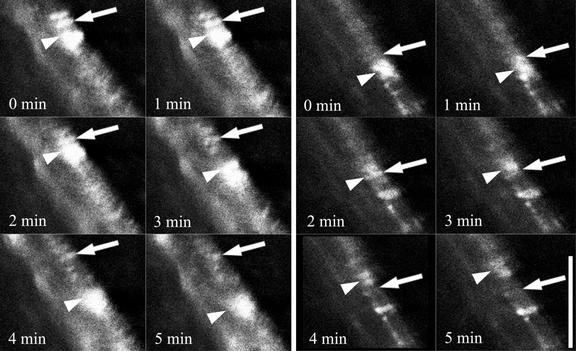
Cellulose synthesis in the developing xylem vessels of Arabidopsis requires three members of the cellulose synthase (CesA) gene family. In young vessels, these three proteins localize within the cell, whereas in older vessels, all three CesA proteins colocalize with bands of cortical microtubules that mark the sites of secondary cell wall deposition. In the absence of one subunit, however, the remaining two subunits are retained in the cell, demonstrating that all three CesA proteins are required to assemble a functional complex. CesA proteins with altered catalytic activity localize normally, suggesting that cellulose synthase activity is not required for this localization. Cortical microtubule arrays are required continually to maintain normal CesA protein localization. By contrast, actin microfilaments do not colocalize with the CesA proteins and are unlikely to play a direct role in their localization. Green fluorescent protein-tagged CesA reveals a novel process in which the structure and/or local environment of the cellulose synthase complex is altered rapidly.


Figures

Figure 1.
Figure 1.
Localization of α-Tubulin and CesA Proteins in Developing Xylem Vessels. Confocal images showing the localization of α-tubulin (left), CesA proteins (middle), and their overlap in distribution (right) in developing vessels from wild-type Arabidopsis roots. IRX3 images were taken from a single root and show early stages of xylem development (top) through the latter stages (bottom). IRX1 and IRX5 images were taken at the latter stages of vessel development. Bar = 25 μm.
Figure 2.
Figure 2.
Localization of α-Tubulin, IRX1, IRX3, and IRX5 in Developing Vessels of irx3-1 and irx5-1 Roots. Confocal images showing the localization of α-tubulin (left), CesA proteins (middle), and their overlap in distribution (right) in developing vessels from irx3-1 (top) and irx5-1 (bottom) Arabidopsis roots. Bar = 25 μm.
Figure 3.
Figure 3.
Localization of α-Tubulin and IRX1, IRX3, and IRX5 in Developing Vessels of irx1-1 Roots. Confocal images showing the localization of α-tubulin (left), CesA proteins (middle), and their overlap in distribution (right) in developing vessels from irx1-1 Arabidopsis roots. Bar = 25 μm.
Figure 4.
Figure 4.
Localization of BiP and IRX1 in Developing Xylem Vessels. Confocal images showing BiP (left), IRX1 (middle), and their degree of colocalization (right) in young (top) and older (middle and bottom) developing xylem vessels from wild-type (WT; top and middle) and irx3-1 (bottom) Arabidopsis roots. Bar = 25 μm.
Figure 5.
Figure 5.
Localization of Actin Microfilaments and IRX3 in Wild-Type Arabidopsis Roots. Confocal images showing actin (left), IRX3 (middle), and their degree of colocalization (right) in developing xylem vessels from wild-type Arabidopsis roots. Bar = 25 μm.
Figure 6.
Figure 6.
Effect of Microtubule Depolymerization on IRX3 Localization. Confocal images showing the localization of α-tubulin (left), IRX3 (middle), and their overlap in distribution (right) in developing vessels from wild-type Arabidopsis roots treated with 10 μM oryzalin for 15, 30, or 60 min or with 0.1% acetone for 60 min. Bar = 25 μm.
Figure 7.
Figure 7.
Localization of IRX3-GFP Fusion in Xylem Vessels. Confocal images showing the distribution of IRX1, IRX5, and IRX3-GFP in roots of irx3-1 plants complemented with an IRX3-GFP construct. IRX1 and IRX5 were visualized using immunofluorescence, whereas IRX3 was visualized using immunofluorescence (IRX3) or directly using GFP fluorescence (IRX3-GFP). Bar = 25 μm.
Figure 8.
Figure 8.
Rapid Alteration in IRX3-GFP Fluorescence during Xylem Development. Two series of successive confocal images taken at 1-min intervals showing IRX3-GFP fluorescence in a developing xylem vessel. Arrows indicate a banded area of IRX3-GFP fluorescence that transiently exhibits a dramatic increase in brightness. Arrowheads indicate a region of IRX3-GFP fluorescence moving along a cell that transiently associates with the stable band of IRX3-GFP fluorescence. Bar = 15 μm.

Similar articles

Cited by

References

    1. Amor, Y., Haigler, C.H., Johnson, S., Wainscott, M., and Delmer, D.P. (1995). A membrane-associated form of sucrose synthase and its potential role in synthesis of cellulose and callose in plants. Proc. Natl. Acad. Sci. USA 92, 9353–9357. - PMC - PubMed
    1. Anthony, R.G., Waldin, T.R., Ray, J.A., Bright, S.W.J., and Hussey, P.J. (1998). Herbicide resistance caused by spontaneous mutation of the cytoskeletal protein tubulin. Nature 393, 260–263. - PubMed
    1. Arioli, T., et al. (1998). Molecular analysis of cellulose biosynthesis in Arabidopsis. Science 279, 717–720. - PubMed
    1. Baskin, T.I. (2001). On the alignment of cellulose microfibrils by cortical microtubules: A review and a model. Protoplasma 215, 150–171. - PubMed
    1. Brower, D.L., and Hepler, P.K. (1976). Microtubules and secondary wall deposition in xylem: The effects of isopropyl N-phenylcarbamate. Protoplasma 87, 91–111. - PubMed

Publication types

MeSH terms

LinkOut - more resources