. 2003 Aug;15(8):1781-94.
doi: 10.1105/tpc.013342.

An Arabidopsis mutant resistant to thaxtomin A, a cellulose synthesis inhibitor from Streptomyces species

Affiliations

An Arabidopsis mutant resistant to thaxtomin A, a cellulose synthesis inhibitor from Streptomyces species

Wolf-Rüdiger Scheible et al. Plant Cell. 2003 Aug.

Abstract

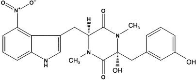
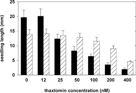
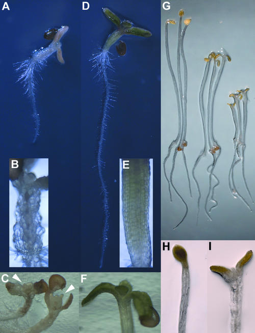
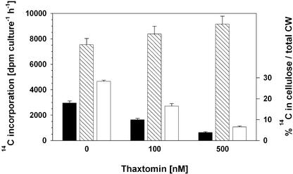
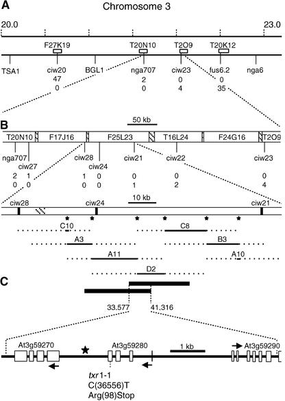
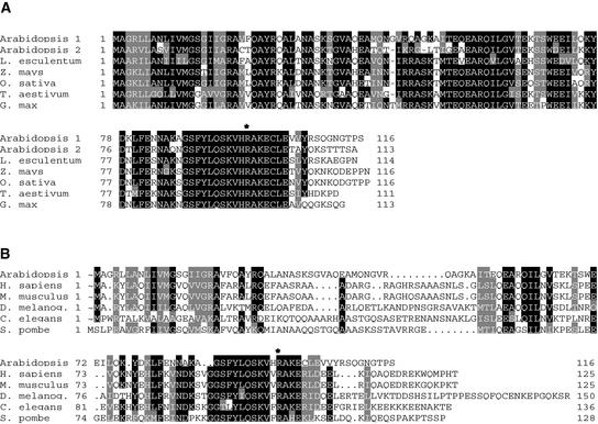
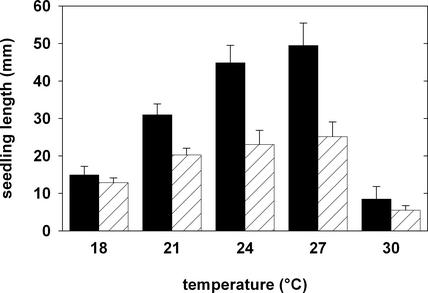
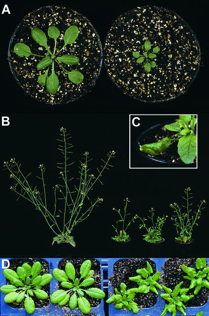
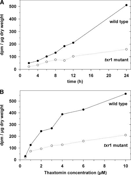
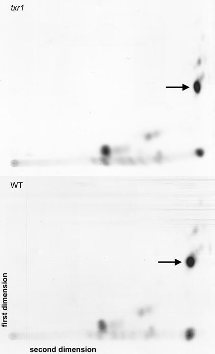
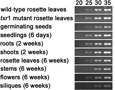
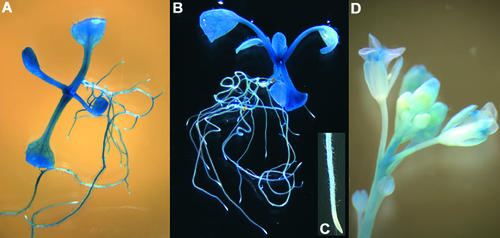
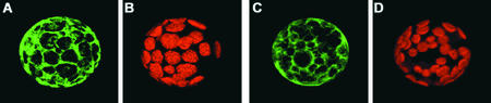
Thaxtomin A is a phytotoxin produced by Streptomyces scabies and other Streptomyces species, the causative agents of common scab disease in potato and other taproot crops. At nanomolar concentrations, thaxtomin causes dramatic cell swelling, reduced seedling growth, and inhibition of cellulose synthesis in Arabidopsis. We identified a mutant of Arabidopsis, designated txr1, that exhibits increased resistance to thaxtomin as a result of a decrease in the rate of toxin uptake. The TXR1 gene was identified by map-based cloning and found to encode a novel, small protein with no apparent motifs or organelle-targeting signals. The protein, which has homologs in all fully sequenced eukaryotic genomes, is expressed in all tissues and during all developmental stages analyzed. Microarray transcript profiling of some 14,300 genes revealed two stomatin-like genes that were expressed differentially in the txr1 mutant and the wild type. We propose that TXR1 is a regulator of a transport mechanism.


Figures

Figure 1.
Figure 1.
Structure of Thaxtomin A.
Figure 2.
Figure 2.
Seedling Length as a Measure of Growth of the Wild Type and the txr1 Mutant (Line BF1) in the Presence of Various Concentrations of Thaxtomin. Each bar represents the mean ± sd of the lengths (measured from the apical meristem to the root tip) of 30 seedlings. Seedlings were grown in Percival chambers on vertical agar plates for 5 days. Wild-type data are shown with black bars, and mutant data are shown with cross-hatched bars.
Figure 3.
Figure 3.
Phenotypes of the Wild Type and the txr1 Mutant (Line BF1) Grown in the Presence of Thaxtomin. (A) to (F) Phenotypes of wild-type and txr1 mutant seedlings after growth on 100 nM thaxtomin for 6 days. (A) and (D) Aspects of a wild-type seedling (A) and a txr1 mutant seedling (D). (B) and (E) Bright-field magnification of a wild-type hypocotyl with drastically swollen, unordered cells (B) and a txr1 mutant hypocotyl with normal, straight cell files (E). (C) and (F) Aspects of shoot apices in the wild type (C) and the txr1 mutant (F). Wild-type shoot apices display drastic cell swelling and severely disturbed organ formation, whereas the shoot apex of txr1 develops normally. Arrowheads in (C) indicate the location where true leaves normally would emerge. (G) to (I) Phenotypes of dark-grown, 5-day-old wild-type seedlings. (G) Thaxtomin concentrations of 50 nM (middle) and 100 nM (right) clearly inhibit hypocotyl elongation compared with seedlings that grew in the absence of the phytotoxin (left). Root elongation is unaffected under these conditions. (H) Etiolated seedlings grown in the absence of thaxtomin have slim hypocotyls and closed cotyledons. (I) Etiolated seedlings grown with 100 nM thaxtomin show swelling symptoms in the upper part of the hypocotyls and the petioles of the cotyledons, forcing the cotyledons to open.
Figure 4.
Figure 4.
Thaxtomin Inhibits 14C-Glucose Incorporation into the Cellulosic Cell Wall Fraction of Dark-Grown Wild-Type Seedlings. A total of 250 4-day-old etiolated seedlings grown in liquid culture were preincubated for 24 h in the presence of 0, 100, or 500 nM thaxtomin and then used for 14C-glucose incorporation assays. Black and cross-hatched bars represent the amount of label incorporated in the acid-insoluble (cellulosic) cell wall (CW) fraction and the acid-soluble cell wall fraction, respectively. White bars indicate the percentage of label in the cellulose fraction relative to the amount of total label in the cell wall (right ordinate). Each bar shows the mean ± se of six independent measurements. Seedling fresh weights in the liquid cultures after preincubation were 170 ± 15 mg, 142 ± 9 mg, and 108 ± 6 mg (mean ± se; n = 6) in the absence of thaxtomin, the presence of 100 nM thaxtomin, or the presence of 500 nM thaxtomin, respectively.
Figure 5.
Figure 5.
Identification of TXR1 by Recombinational Mapping and Cosmid Complementation. (A) Representation of the bottom part of Arabidopsis chromosome 3 showing a megabase scale, the positions of cleaved amplified polymorphic sequence markers TSA1 and BGL1, and the positions of SSLP markers nga6, nga707, ciw20, ciw23, and fus6.2. AGI BAC clones containing the SSLP markers used for fine mapping are represented by white boxes. The number of recombination events (meiotic breakpoints) found for each marker in 2304 chromosomes examined is given. (B) Close-up of the region between flanking SSLP markers nga707 and ciw23 showing the names, the nonoverlapping parts (open boxes) and overlapping parts (cross-hatched boxes) of BAC clones, and the positions and numbers of recombinant events found for five additional ciw SSLP markers. The enlarged view of the region between ciw28 and ciw21 depicts the positions of probes (asterisks) used for cosmid library screening and the positions of seven cosmid clones covering the 83.4-kb interval. Dotted lines indicate the regions in which the cosmid clones end. The exact positions and sizes, as well as the 7.7-kb overlap of the two txr1 complementing cosmids, A11 and D2, on BAC clone F25L23 are shown below. (C) Representation of the predicted genes within the 7.7-kb segment. The point mutation in the txr1-1 allele and the introduced stop codon are shown. Arrows indicate the direction of gene transcription and are positioned at the translational start codons.
Figure 6.
Figure 6.
Amino Acid Alignment of TXR1 Homologs. Black boxes indicate regions in which at least five of seven (A) or five of six (B) residues are identical, and gray boxes indicate conserved residues. The position of the stop codon in TXR1 is marked with an asterisk. (A) Alignment of a selection of homologs from monocotyledonous and dicotyledonous plants as deduced from EST sequences (tomato [Lycopersicon esculentum], maize [Zea mays], wheat [Triticum aestivum], and soybean [Glycine max]) or genomic sequences (Arabidopsis and rice [Oryza sativa]). TXR1 is designated Arabidopsis 1. (B) Alignment of TXR1 with eukaryotic sequences from human (Homo sapiens), mouse (Mus musculus), fruit fly (Drosophila melanogaster), worm (Caenorhabditis elegans), and fission yeast (Schizosaccharomyces pombe).
Figure 7.
Figure 7.
Growth of Wild-Type and txr1 Mutant Seedlings in Response to Different Temperatures. Each bar represents the mean ± sd of the lengths (measured from the apical meristem to the root tip) of 20 seedlings grown in Percival chambers on vertical agar plates for 7 days. Wild-type data are shown with black bars, and mutant data are shown with cross-hatched bars.
Figure 8.
Figure 8.
Growth Phenotypes of txr1 Mutants during Later Stages of Development. (A) and (B) Wild-type (left) and txr1 mutant (right) plants grown for 4 weeks (A) or 6 weeks (B) on soil under greenhouse conditions. (C) Characteristic rosette leaf shape of a 5-week-old txr1 mutant. (D) Wild-type (left) and txr1 mutant (right) plants grown for 7 weeks on soil in a 12-h-light/12-h-dark cycle at 19/16°C. The leaf phenotype of txr1 mutants became visible after ∼5 weeks under these conditions.
Figure 9.
Figure 9.
Uptake of 3H-Thaxtomin by Wild-Type and txr1 Mutant Seedlings. (A) Time-dependent uptake of 3H-thaxtomin by wild-type seedlings (closed symbols) and txr1 mutant seedlings (open symbols) in 2 μM thaxtomin. (B) Concentration-dependent uptake of 3H-thaxtomin by wild-type seedlings (closed symbols) and txr1 mutant seedlings (open symbols). Seedlings were incubated in toxin for 12 h at the indicated concentrations.
Figure 10.
Figure 10.
Two-Dimensional Thin Layer Chromatograms of 3H-Thaxtomin–Labeled Extracts from the txr1 Mutant and the Wild Type. Arrows indicate the positions of thaxtomin A. WT, wild type.
Figure 11.
Figure 11.
Reverse Transcriptase–Mediated PCR Analysis of TXR1 Expression. The level of a 224-bp TXR1-specific PCR product was visualized after 20, 25, 30, and 35 PCR cycles using a first-strand cDNA template from 4-week-old wild-type and mutant rosette leaves, 3-day-old germinating wild-type seeds, 6-day-old wild-type seedlings, roots and shoots from 2-week-old wild-type plants grown on vertical agar plates, and rosette leaves, stems, flowers, and green siliques from 6-week-old adult wild-type plants.
Figure 12.
Figure 12.
Expression of GUS Activity under the Control of a 1.76-kb TXR1 Promoter Fragment. Shown are bright-field (A) and dark-field (B) images of GUS staining in 14-day-old vegetative-stage plantlets as well as a representative root tip of a 14-day-old plantlet (C) and an inflorescence stem of an adult 1-month-old plant (D).
Figure 13.
Figure 13.
Localization of TXR1 Fused at the N and C Termini to GFP in Protoplasts. Green fluorescence of GFP-TXR1 (i.e., GFP is fused to the N terminus of TXR1) (A) and TXR1-GFP (C). (B) and (D) show the chlorophyll fluorescence for the protoplasts shown in (A) and (C), respectively. Both fusion constructs are expressed under the control of the 35S promoter of Cauliflower mosaic virus.

References

    1. Acuna, I.A., Strobel, G.A., Jacobsen, B.L., and Corsini, D.L. (2001). Glucosylation as a mechanism of resistance to thaxtomin A in potatoes. Plant Sci. 161, 77–88.
    1. Anzai, H., Yoneyama, K., and Yamagushi, I. (1989). Transgenic tobacco resistant to a bacterial disease by the detoxification of a pathogenic toxin. Mol. Gen. Genet. 219, 492–494.
    1. Bukhalid, R.A., Takeuchi, T., Labeda, D., and Loria, R. (2002). Horizontal transfer of the plant virulence gene, nec1, and flanking sequences among genetically distinct strains in the diastatochromogenes cluster. Appl. Environ. Microbiol. 68, 738–744. - PMC - PubMed
    1. Clough, S.J., and Bent, A.F. (1998). Floral dip: A simplified method for Agrobacterium-mediated transformation of Arabidopsis thaliana. Plant J. 16, 735–743. - PubMed
    1. Cutler, S.R., Ehrhardt, D.W., Griffitts, J.S., and Somerville, C.R. (2000). Random GFP::cDNA fusions enable visualization of subcellular structures in cells of Arabidopsis at high frequency. Proc. Natl. Acad. Sci. USA 97, 3718–3723. - PMC - PubMed

Publication types

MeSH terms