. 2003 Aug;15(8):1872-87.
doi: 10.1105/tpc.012427.

Arabidopsis AtGPAT1, a member of the membrane-bound glycerol-3-phosphate acyltransferase gene family, is essential for tapetum differentiation and male fertility

Affiliations

Arabidopsis AtGPAT1, a member of the membrane-bound glycerol-3-phosphate acyltransferase gene family, is essential for tapetum differentiation and male fertility

Zhifu Zheng et al. Plant Cell. 2003 Aug.

Abstract

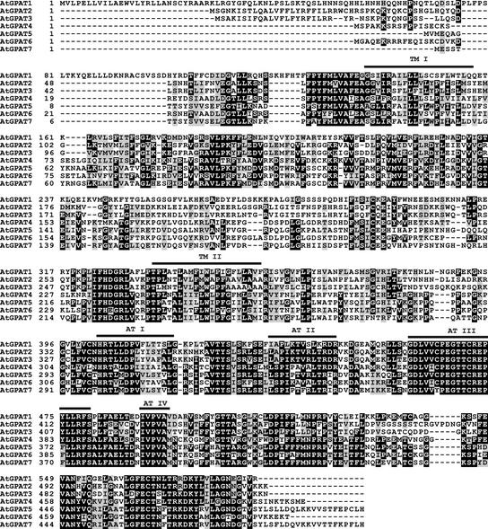
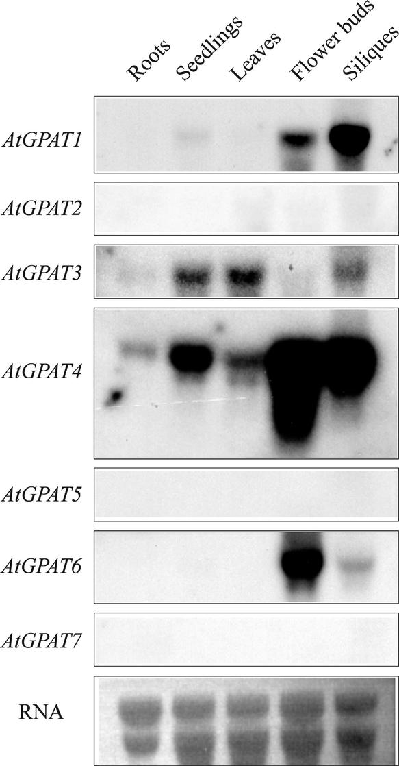
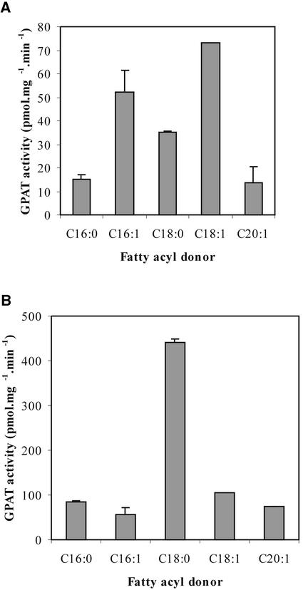
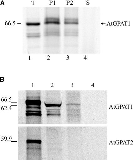
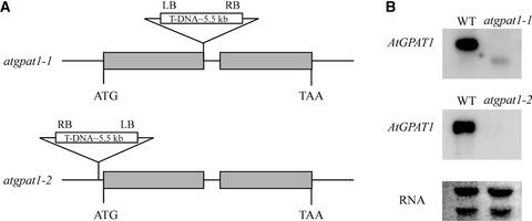
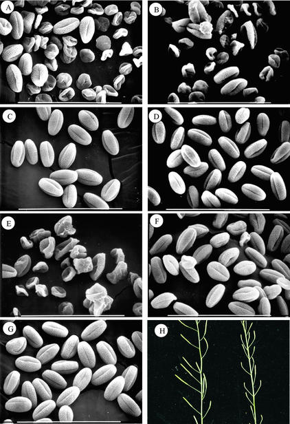
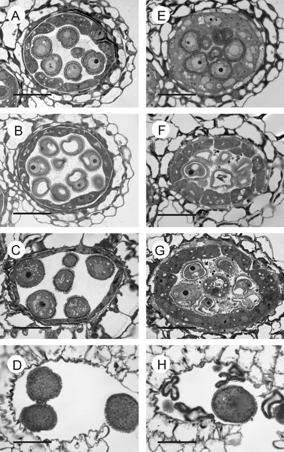
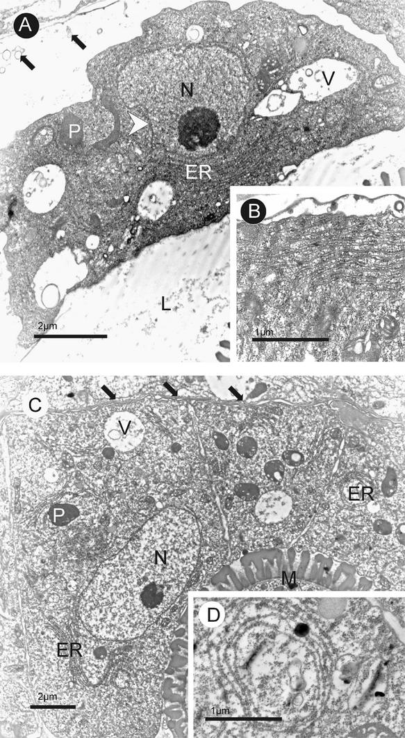
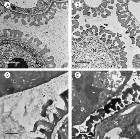
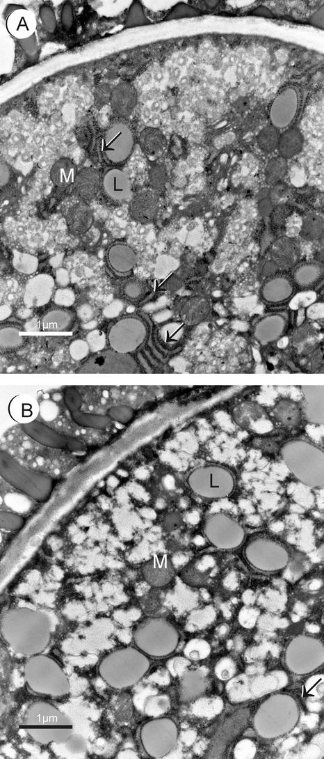
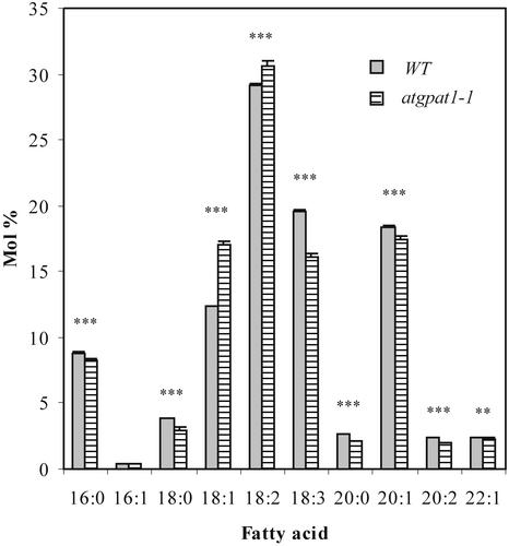
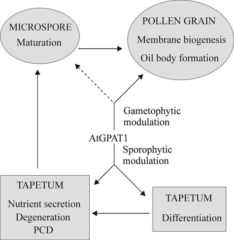
Membrane-bound glycerol-3-phosphate acyltransferase (GPAT; EC 2.3.1.15) mediates the initial step of glycerolipid biosynthesis in the extraplastidic compartments of plant cells. Here, we report the molecular characterization of a novel GPAT gene family from Arabidopsis, designated AtGPAT. The corresponding polypeptides possess transmembrane domains and GPAT activity when expressed heterologously in a yeast lipid mutant. The functional significance of one isoform, AtGPAT1, is the focus of the present study. Disruption of the AtGPAT1 gene causes a massive pollen development arrest, and subsequent introduction of the gene into the mutant plant rescues the phenotype, illustrating a pivotal role for AtGPAT1 in pollen development. Microscopic examinations revealed that the gene lesion results in a perturbed degeneration of the tapetum, which is associated with altered endoplasmic reticulum profiles and reduced secretion. In addition to the sporophytic effect, AtGPAT1 also exerts a gametophytic effect on pollen performance, as the competitive ability of a pollen grain to pollinate is dependent on the presence of an AtGPAT1 gene. Deficiency in AtGPAT1 correlates with several fatty acid composition changes in flower tissues and seeds. Unexpectedly, however, a loss of AtGPAT1 causes no significant change in seed oil content.


Figures

Figure 1.
Figure 1.
Deduced Amino Acid Sequence Alignment of the Members of the AtGPAT Family in Arabidopsis. Four conserved acyltransferase motifs are indicated above the alignment (AT I, AT II, AT III, and AT IV). Two transmembrane domains (TM I and TM II) were predicted computationally. Identical and similar amino acid residues are shaded black and gray, respectively.
Figure 2.
Figure 2.
Expression Analysis of AtGPAT Genes. Approximately 10 μg of total RNA in each lane was used for hybridization with the individual AtGPAT probes. Equal RNA loading was verified by ethidium bromide staining. The autoradiographs were generated by exposure for 24 h.
Figure 3.
Figure 3.
Fatty Acyl Specificity of AtGPAT1. Assays of GPAT were conducted with 14C–G-3-P as described in Methods in the absence (A) and presence (B) of EDTA. C16:0, palmitoyl-CoA; C16:1, palmitoleoyl-CoA; C18:0, stearoyl-CoA; C18:1, oleoyl-CoA; C20:1, eicosenoic acyl-CoA.
Figure 4.
Figure 4.
Subcellular Targeting of AtGPAT1. AtGPAT1 was synthesized in vitro using transcription/translation systems with 35S-Met. (A) Integration of AtGPAT1 into ER-derived membranes. Lane 1, total translation products (T); lane 2, microsomal pellet (P1) of the translation mixture after the high-salt wash; lane 3, microsomal pellet (P2) derived from the translation mixture washed with the high-salt solution followed by alkaline extraction; lane 4, supernatant (S) generated from the alkaline extraction of the high-salt-washed microsomes precipitated with trichloroacetic acid. The sizes of the synthesized polypeptides are shown at left in kilodaltons. (B) In vitro import of AtGPAT1 into pea mitochondria. Lane 1, total translation products; lane 2, labeled polypeptides imported into mitochondria; lanes 3 and 4, the imported polypeptides treated with proteinase K in the absence and presence, respectively, of 0.5% (v/v) Triton X-100. The sizes of the nonprocessed and processed proteins are indicated in kilodaltons at left. AtGPAT2 was used as a control.
Figure 5.
Figure 5.
Genetic Analysis of the atgpat1-1 and atgpat1-2 Mutants. (A) Genomic organization of the atgpat1-1 and atgpat1-2 loci. Gray horizontal boxes represent exons. The T-DNA insert is not drawn to scale. LB, T-DNA left border; RB, T-DNA right border. (B) RNA gel blot analysis of AtGPAT1 transcript in the wild type (WT) and mutants. Approximately 10 μg of total RNA from siliques was loaded in each lane.
Figure 6.
Figure 6.
Male Fertility in Wild-Type and Mutant Plants. (A) to (G) Scanning electron micrographs of mature pollen grains from atgpat1-1 homozygous (A) and heterozygous (C) plants, atgpat1-2 homozygous (B) and heterozygous (D) plants, atgpat1-1 homozygous plants transformed with the control vector pRD400 alone (E) and with the vector carrying the AtGPAT1 gene (F), and a wild-type plant (G). Bars = 100 μm. (H) Silique development in atgpat1-1 homozygous plants transformed with the AtGPAT1 gene (left) and the control vector (right).
Figure 7.
Figure 7.
Light Micrographs of Cross-Sections of Wild-Type and atgpat1-1 Anthers. The left and right micrographs are from wild-type and atgpat1-1 anthers, respectively. Stages are defined according to Owen and Makaroff (1995). Bars = 20 μm. (A) and (E) Microspore-release stage. (B) and (F) Ring-vacuolated microspore stage. (C) and (G) Bicellular pollen grain stage. (D) and (H) Mature pollen grain stage.
Figure 8.
Figure 8.
Transmission Electron Micrographs of Cross-Sections of Wild-Type and atgpat1-1 Anthers at Microspore-Release Stage II. (A) and (B) Wild-type anthers. (C) and (D) atgpat1-1 anthers. The tapetal cell wall is completely dissolved in the wild type (A) but remains intact in the mutant (C), as indicated by black arrows. The white arrowhead shows dented membranes in the nucleus of the wild-type tapetum in (A). Insets (B) and (D) show ER stack and ER ring structures in wild-type and mutant tapetal cells, respectively. ER, endoplasmic reticulum; L, locule; M, microspore; N, nucleus; P, plastid; V, vacuole.
Figure 9.
Figure 9.
Secretion Processes in Wild-Type and atgpat1-1 Tapeta. Transmission electron micrographs of cross-sections of wild-type ([A] and [C]) and atgpat1-1 ([B] and [D]) anthers. At the ring-vacuolated microspore stage ([A] and [B]), a high concentration of fibrillar material is distributed uniformly throughout the wild-type locule (A), whereas much less material appears in the mutant locule (B). At the early bicellular stage ([C] and [D]), many osmophilic material–containing vesicles are secreted from the wild-type tapetum into the locule (C), whereas fewer vesicles are present in the mutant locule (D). E, elaioplast; F, fibrillar material; L, lipid body; N, nucleus; pg, pollen grain; T, tapetal cell; V, vesicle.
Figure 10.
Figure 10.
Transmission Electron Micrographs of Mature Pollen Grains from Wild-Type and atgpat1-1 Plants. (A) Wild-type pollen grain. (B) atgpat1-1 pollen grain. Arrows indicate ER cisternae. L, lipid body; M, mitochondria.
Figure 11.
Figure 11.
Seed Fatty Acid Compositions of Wild-Type and Mutant Plants Grown under Identical Conditions. Only the fatty acid composition of atgpat1-1 is shown because the two mutant lines have similar fatty acid profiles. Results shown are means ± se from five independent assays. Data were analyzed with paired t tests for individual fatty acids. Double asterisks indicate P < 0.01; triple asterisks indicate P < 0.001. WT, wild type.
Figure 12.
Figure 12.
Involvement of AtGPAT1 in Tapetal Differentiation and Pollen Development. The placement of solid arrows is based on evidence presented in this work. The dashed arrow denotes a potential role for AtGPAT1. PCD, programmed cell death.

References

    1. Altschul, S.F., Gish, W., Miller, W., Myers, E.W., and Lipman, D.J. (1990). Basic local alignment search tool. J. Mol. Biol. 215, 403–410. - PubMed
    1. Athenstaedt, K., and Daum, G. (1999). Phosphatidic acid, a key intermediate in lipid metabolism. Eur. J. Biochem. 266, 1–16. - PubMed
    1. Bafor, M., Stobart, A.K., and Stymne, S. (1990). Properties of the glycerol acylating enzyme in microsomal preparations from the developing seeds of safflower (Carthamus tinctorius) and turnip rape (Brassica campestris) and their ability to assemble cocoa-butter type fats. J. Am. Oil Chem. Soc. 67, 217–225.
    1. Balk, J., and Leaver, C.J. (2001). The PET1-CMS mitochondrial mutation in sunflower is associated with premature programmed cell death and cytochrome c release. Plant Cell 13, 1803–1818. - PMC - PubMed
    1. Bligh, E.G., and Dyer, W.J. (1959). A rapid method of total lipid extraction and purification. Can. J. Biochem. Physiol. 37, 911–917. - PubMed

Publication types

MeSH terms