. 2003 Aug;15(8):1904-17.
doi: 10.1105/tpc.013995.

Regulation and processing of maize histone deacetylase Hda1 by limited proteolysis

Affiliations

Regulation and processing of maize histone deacetylase Hda1 by limited proteolysis

Alexandra Pipal et al. Plant Cell. 2003 Aug.

Abstract

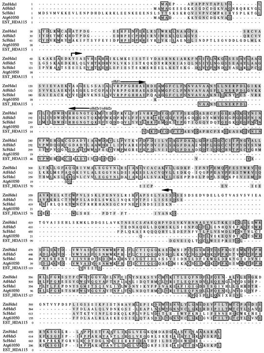
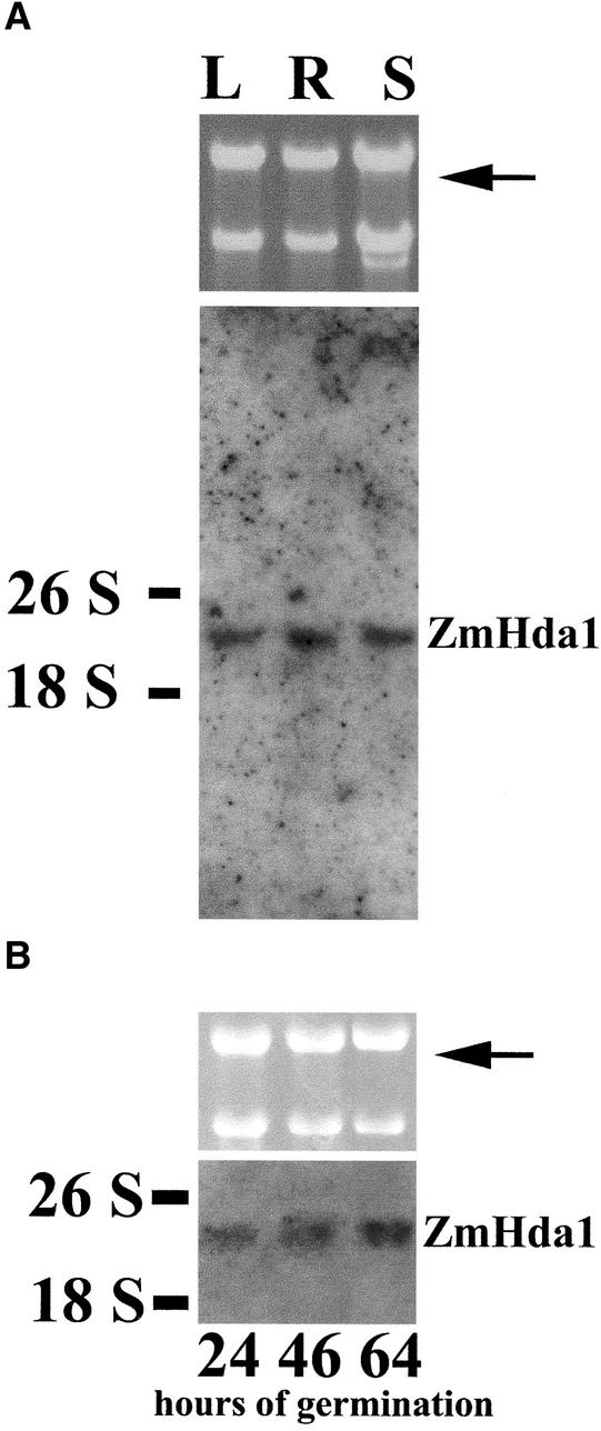
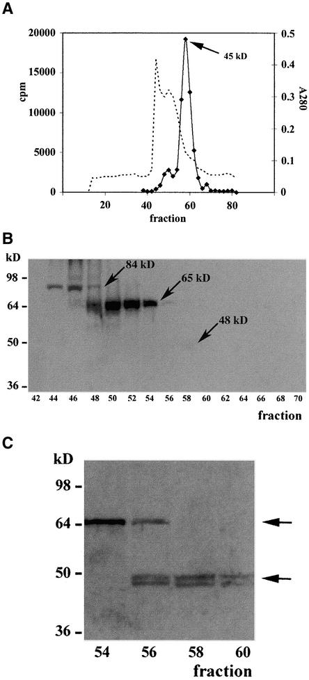
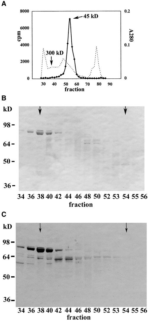
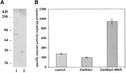
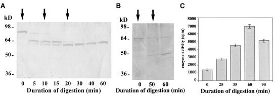
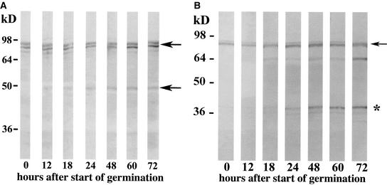
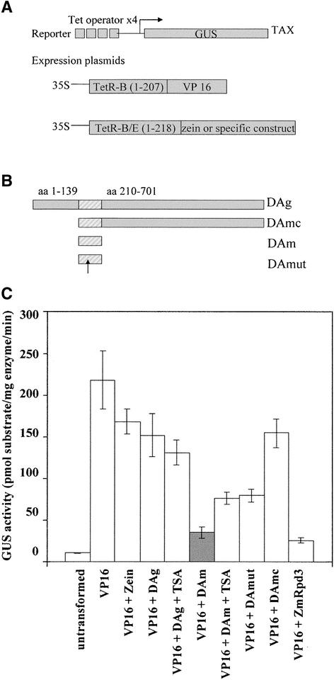
A maize histone deacetylase gene was identified as a homolog of yeast Hda1. The predicted protein corresponds to a previously purified maize deacetylase that is active as a protein monomer with a molecular weight of 48,000 and is expressed in all tissues of germinating embryos. Hda1 is synthesized as an enzymatically inactive protein with an apparent molecular weight of 84,000 that is processed to the active 48-kD form by proteolytic removal of the C-terminal part, presumably via a 65-kD intermediate. The enzymatically inactive 84-kD protein also is part of a 300-kD protein complex of unknown function. The proteolytic cleavage of ZmHda1 is regulated during maize embryo germination in vivo. Expression of the recombinant full-length protein and the 48-kD form confirmed that only the smaller enzyme form is active as a histone deacetylase. In line with this finding, we show that the 48-kD protein is able to repress transcription efficiently in a reporter gene assay, whereas the full-length protein, including the C-terminal part, lacks full repression activity. This report on the processing of Hda1-p84 to enzymatically active Hda1-p48 demonstrates that proteolytic cleavage is a mechanism to regulate the function of Rpd3/Hda1-type histone deacetylases.


Figures

Figure 1.
Figure 1.
Comparison of the Amino Acid Sequences of ZmHda1 and Related Proteins. ZmHda1 was aligned with the corresponding amino acid sequences of S. cerevisiae (ScHda1), two proteins of Arabidopsis (AtHda5 and Atg61050), and a maize protein (EST HDA115). Identical amino acids are boxed, and homologous amino acids are shaded. The positions of the oligonucleotide primers for the initial amplification of a ZmHda1 fragment are indicated. The area of peptides derived from protein microsequencing of ZmHda1-p48 (the former HD1A) is marked with arrows.
Figure 2.
Figure 2.
Expression of ZmHda1 mRNA in Different Maize Organs and at Various Germination Stages of Embryos. Total RNA was isolated from leaf (L), root (R), and shoot (S) of plant seedlings at 72 h after the start of embryo germination (A) or at 24, 46, and 64 h after the start of embryo germination (B). RNA was subjected to agarose gel electrophoresis with subsequent RNA gel blot analysis. The blot was hybridized with a ZmHda1-specific, digoxigenin-labeled, 513-bp DNA fragment. An ethidium bromide–stained gel is shown as an input control (top gels; 18 and 26 S RNA). The arrows indicate the positions of ZmHda1 mRNA.
Figure 3.
Figure 3.
Immunological Analysis of Partially Purified ZmHda1. (A) Proteins of the soluble supernatant from chromatin preparations of maize embryos at 72 h after the start of germination were separated by Q-Sepharose Big Beads chromatography. Fractions containing histone deacetylase activity were pooled and subjected to S75 gel filtration chromatography. The bulk of enzyme activity eluted in a peak at an apparent molecular weight of ∼45,000. The dashed line depicts the A280 as a rough estimate of the protein concentration. (B) Fractions from the S75 gel filtration chromatography were blotted onto a nitrocellulose membrane, and ZmHda1 bands were detected with anti-ZmHda1 antibodies (peptide antibodies). Prominent bands appeared at molecular weights of 84,000 and 65,000 (p84 and p65; arrows). In fraction 58, a very faint band was visible in the original wet blot at a molecular weight of ∼48,000 (p48; arrow). (C) Chromatographic fractions (54 to 60) from S75 gel filtration chromatography were concentrated by trichloroacetic acid precipitation, subjected to SDS-PAGE with subsequent blotting onto a nitrocellulose membrane, and immunodetected with anti-ZmHda1 peptide antibodies. ZmHda1-p48 appears exactly in the histone deacetylase activity peak (see [A]). Arrows mark the positions of ZmHda1-p65 and ZmHda1-p48.
Figure 4.
Figure 4.
Immunological Analysis of Partially Purified ZmHda1 after S200 Gel Filtration Chromatography. (A) The soluble supernatant from a chromatin preparation of 500 g of maize embryos at 72 h after the start of germination was subjected to Q-Sepharose Big Beads chromatography. Those fractions containing ZmHda1 activity without contamination by ZmRpd3 were pooled and subjected to S200 gel filtration chromatography. ZmHda1 enzyme activity eluted in a peak at an apparent molecular weight of ∼45,000. The dashed line depicts the A280 as a rough estimate of the protein concentration. (B) Fractions from the S200 gel filtration chromatography were blotted onto a nitrocellulose membrane, and proteins were immunodetected with anti-ZmHda1 antibodies (peptide antibodies). Arrows in fractions 38 and 54 mark the molecular weights of 300,000 (ZmHda1-p84) and 45,000 (ZmHda1-p48), respectively. (C) The same blot shown in (B) was immunoblotted with antibodies against the C-terminal part of ZmHda1. Arrows in fractions 38 and 54 mark the molecular weights of 300,000 (ZmHda1-p84) and 45,000 (ZmHda1-p48), respectively.
Figure 5.
Figure 5.
Expression of ZmHda1-p84 and ZmHda1-p48 in Insect Cells Using the Baculovirus Expression System. (A) SDS-PAGE of recombinant, His-tagged, full-length ZmHda1-p84 (amino acids 4 to 701; lane 1) and ZmHda1-p48 lacking the C-terminal 272 amino acids (amino acids 23 to 429; lane 2). Insect cell extracts were subjected to SDS-PAGE and immunoblotted using anti-ZmHda1 peptide antibodies. (B) Histone deacetylase activity of recombinant ZmHda1-p84 and ZmHda1-p48. The control bar indicates histone deacetylase activity bound to Talon metal affinity resin of nontransfected insect cells (background activity); the ZmHda1 bar indicates histone deacetylase activity of insect cells transfected with full-length p84; the ZmHda1-48kD bar indicates histone deacetylase activity of insect cells transfected with the C-terminally truncated form.
Figure 6.
Figure 6.
In Vitro Conversion of ZmHda1-p84 to Smaller Enzyme Forms by Carboxypeptidase Treatment. (A) Chromatographic fractions (pool of 44 and 45; see Figure 3A) containing ZmHda1-p84 were digested with carboxypeptidase (30 units) for 60 min. The digestion process was monitored by SDS-PAGE at the indicated times. Arrows mark the addition of carboxypeptidase. The gel was blotted onto a nitrocellulose membrane, and ZmHda1 was immunodetected with anti-ZmHda1 peptide antibodies. (B) Chromatographic fractions (pool of 52 and 53; see Figure 3A) containing ZmHda1-p65 were digested with carboxypeptidase for 60 min. (C) Histone deacetylase activity was measured during 90 min of carboxypeptidase digestion of chromatographic fractions 52 and 53 (see Figure 3A) containing ZmHda1-p65.
Figure 7.
Figure 7.
Immunological Characterization of ZmHda1 Forms during Maize Embryo Germination. (A) Embryos were harvested into liquid nitrogen at various times during embryo germination (hours after the start of imbibition). Proteins were extracted, and equal amounts of protein were subjected to SDS-PAGE. The gel was blotted onto a nitrocellulose membrane, and proteins were immuno- detected with anti-ZmHda1 antibodies (peptide antibodies). Arrows mark the positions of the high molecular weight ZmHda1 form and the 48-kD form. (B) The same blot shown in (A) was immunoblotted with antibodies against the C-terminal part of ZmHda1. The arrow marks the position of the high molecular weight ZmHda1 form, and the asterisk marks the position of the cleaved C-terminal peptide.
Figure 8.
Figure 8.
Effect of ZmHda1 Constructs on the Repression of Gene Transcription in Tobacco Protoplasts. (A) Scheme of the reporter and expression plasmids used in the tobacco protoplast. The tobacco TAX line contains a reporter cassette that is integrated stably in the genome and allows for the expression of a reporter GUS controlled by a minimal promoter and four consecutive upstream TetR binding sites. Cotransfection of two plasmids with the 35S promoter driving the expression of the VP16 transcriptional activator and the protein of interest, respectively, both fused to the DNA binding domain of TetR, results in the competition of the two fusion proteins for the TET binding sites. The effect on transcription is monitored by determining GUS activity levels (Bohner et al., 1999). (B) ZmHda1 constructs. The striped area represents the core catalytic histone deacetylase domain. The arrow depicts the position of the mutated His in the DAmut construct. aa, amino acids. (C) Full-length ZmHda1 or ZmHda1 derivatives fused to the DNA binding domain of TetR were used together with plasmid expressing a TetR fusion of VP16 to transform protoplasts prepared from the tobacco TAX line. The effect on transcription was determined by measuring the activity of the GUS reporter gene. GUS activity is reported as pmol of 7-OH-4-methylcoumarin per milligram of protein per minute. “Untransformed” indicates protoplast samples transformed with the DNA carrier only. Error bars indicate the standard errors of assays performed on six independent transfections.

Similar articles

Cited by

References

    1. Bertos, N.R., Wang, A.H., and Yang, Y.J. (2001). Class II histone deacetylases: Structure, functions, and regulations. Biochem. Cell Biol. 79, 243–252. - PubMed
    1. Bohner, S., Lenk, I.I., Rieping, M., Herold, M., and Gatz, C. (1999). Technical advances: Transcriptional activator TGV mediates dexamethasone-inducible and tetracycline-inactivable gene expression. Plant J. 19, 87–95. - PubMed
    1. Borchers, C., Peter, J.F., Hall, M.C., Kunkel, T.A., and Tomer, K.B. (2000). Identification of in-gel digested proteins by complementary peptide mass fingerprinting and tandem mass spectrometry data obtained on an electrospray ionization quadrupole time-of-flight mass spectrometer. Anal. Chem. 72, 1163–1168. - PubMed
    1. Boyer, L.A., Logie, C., Bonte, E., Becker, P.B., Wade, P.A., Wolffe, A.P., Wu, C., Imbalzano, A.N., and Peterson, C.L. (2000). Functional delineation of three groups of the ATP-dependent family of chromatin remodeling enzymes. J. Biol. Chem. 275, 18864–18870. - PubMed
    1. Bradford, M.M. (1976). A rapid and sensitive method for the quantitation of microgram quantities of protein utilizing the principle of protein-dye binding. Anal. Biochem. 72, 248–254. - PubMed

Publication types

MeSH terms

LinkOut - more resources