. 2003 Aug;4(8):813-8.
doi: 10.1038/sj.embor.embor902. Epub 2003 Jul 25.

Integrin-alpha3 mediates binding of Chordin to the cell surface and promotes its endocytosis

Affiliations

Integrin-alpha3 mediates binding of Chordin to the cell surface and promotes its endocytosis

Juan Larraín et al. EMBO Rep. 2003 Aug.

Abstract

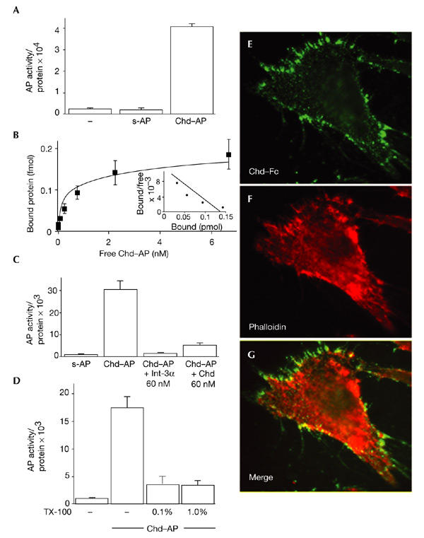
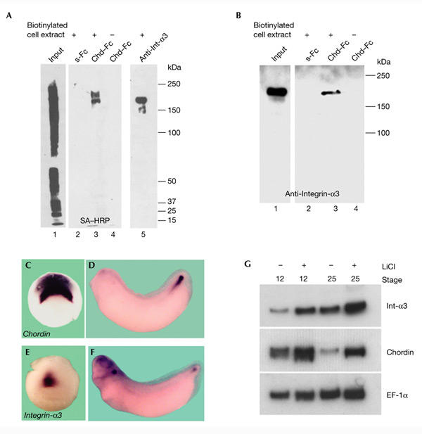
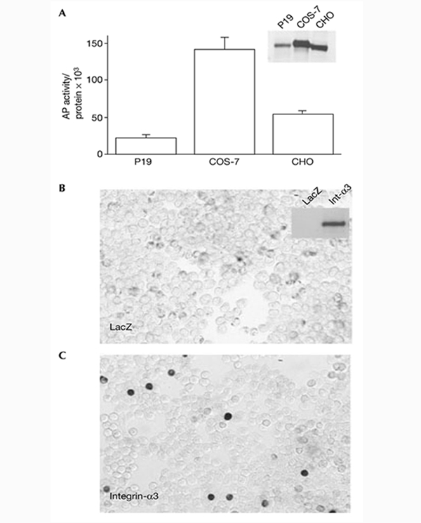
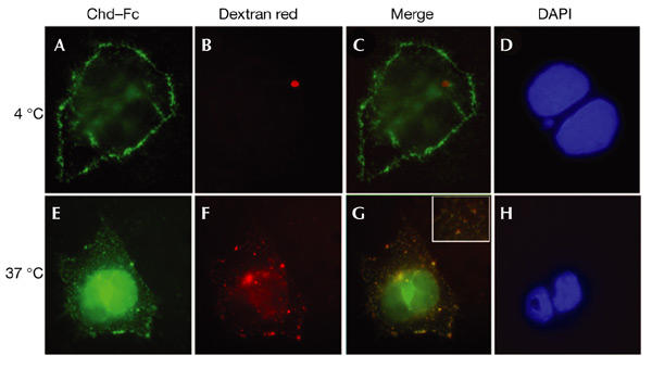
Dorsoventral patterning in animal development is regulated by a morphogenetic gradient of Bone morphogenetic protein signalling, which is established by a set of proteins that are conserved from Drosophila to vertebrates. These include Chordin (Chd)/Short gastrulation, Xolloid/Tolloid and Twisted gastrulation. Here, we report the identification of a cell-surface component of this morphogenetic pathway. Prompted by the observation that Chd protein bound to the surface of certain cell lines with subnanomolar affinity, we identified two cell-surface proteins that bind to Chd, one of which corresponds to Integrin-alpha3. Integrin-alpha3 and Chd are co-expressed in the Xenopus embryo. Transfection of Integrin-alpha3 increased the binding of Chd to the cell surface, which was competed by an excess of soluble Integrin-alpha3. After binding to the cell surface, Chd was translocated into intracellular endocytic compartments in a temperature-dependent manner. We propose that Integrin-alpha3 may regulate the concentration of Chd protein in the extracellular space by endocytosis.


Figures

Figure 1
Figure 1
Chordin binds to the cell surface. (A) Chordin–alkaline-phosphatase (Chd–AP) binds to COS-7 cells. COS-7 cells were incubated with Chd–AP or secreted AP (s-AP) for 2 h at 4 °C, and the amount of activity of bound AP was quantified using a fluorescent AP substrate. Equal loading was confirmed by protein measurements. (B) Binding curve and Scatchard analysis of Chd–AP binding to COS-7 cells. A dissociation constant (Kd) of 0.24 nM was estimated. (C) COS-7 cells were incubated with Chd–AP in the presence of 60 nM recombinant Integrin-α3 (Int-α3; Chemicon) or mouse Chd (R&D Systems), and the amount of bound Chd–AP was quantified using a fluorescent AP substrate. Other cysteine-rich-repeat-containing proteins were not tested because they are not available in purified form. (D) Chd binding requires an intact cell membrane. COS-7 cells were incubated with 0.1% or 1.0% Triton X-100 (TX-100), and Chd–AP was bound and quantified. (E) Chd binding colocalizes with actin filaments. 10T1/2 fibroblasts were incubated with pure Chd–Fc at 4 °C, fixed and permeabilized, and binding was visualized with an anti-human IgG antibody conjugated to Alexa Fluor 486. (F) The same cell as in (E), stained with phalloidin-Alexa558 to visualize actin. (G) Merge of (E) and (F). Note the overlap between Chd–Fc and actin (yellow).
Figure 2
Figure 2
Chordin affinity columns bind biotinylated cell-surface Integrin-α3. (A) A Chordin (Chd)–Fc affinity matrix binds two distinct cell-surface proteins in COS-7 cells. Cell extracts containing biotinylated surface proteins from COS-7 cells were bound to secreted Fc (s-Fc; lane 2) or Chd–Fc (lane 3) columns, or were immunoprecipitated using an anti-Integrin-α3 (Int-α3) antibody (lane 5). Proteins bound to the columns were analysed by immunoblotting with streptavidin–horseradish-peroxidase (SA–HRP; Pierce). Lane 1 shows loading of 1% of the total biotinylated cell lysate. (B) A Chordin affinity matrix binds Integrin-α3. COS-7 cell extracts were bound to s-Fc (lane 2) or Chd–Fc affinity columns (lane 3), eluted, and analysed by immunoblotting with anti-Integrin-α3. (CG) Chordin and Integrin-α3 are co-expressed during embryonic development. In situ hybridization analysis of chordin (C,D) and integrin-a3 (E,F) expression at stages 10.5 and 32. (G) RT–PCR (PCR after reverse transcription) analysis of chordin and integrin-a3 messenger RNA levels in control and LiCl-treated embryos. Elongation factor-1α (EF-1α) was used as a loading control.
Figure 3
Figure 3
Chordin binds to the cell surface through Integrin-α3. (A) Binding of Chordin–alkaline-phosphatase (Chd–AP) to P19, COS-7 and CHO cell lines. The inset shows an anti-Integrin-α3 immunoblot of cell extracts from the three cell lines. The amount of Chd–AP binding correlates with endogenous levels of Integrin-α3. (B,C) Chd–AP binds to 293T cells transfected with integrin-a3. 293T cells were transfected with LacZ or human integrin-a3 full-length complementary DNAs, incubated with recombinant Chd–AP and stained for AP activity. The inset shows an anti-Integrin-α3 (Int-α3) immunoblot of the corresponding cell extracts.
Figure 4
Figure 4
Chordin–Fc translocates into intracellular endocytic compartments at 37 °C. Cells were incubated on ice with Chordin (Chd)–Fc for 2 h, washed, and then incubated with Texas-Red–dextran for 30 min on ice (AD) or at 37 °C (EH). (A,E) Localization of Chd–Fc (green) bound to COS-7 cells. (B,F) Localization of Texas-Red–dextran (red) in punctate endocytic compartments after shifting to 37 °C. (C,G) Merged images. (D,H) 4,6-diamidino-2-phenylindole (DAPI) staining of nuclear DNA. Note that at 4 °C, most of the Chd–Fc is detected at the cell surface, but after 30 min at 37 °C it colocalizes with endocytic vesicles in the cytoplasm that contain Texas-Red–dextran (G). The red spot in (B) is artefactual, but shows that red fluorescence does not bleed into the green fluorescence channel.

Similar articles

Cited by

References

    1. Araujo H., Negreiros E. & Bier E. ( 2003) Integrins modulate Sog activity in the Drosophila wing. Development, in the press. - PubMed
    1. Davis S. et al. . ( 1996) Isolation of angiopoietin-1, a ligand for the TIE2 receptor, by secretion-trap expression cloning. Cell, 87, 1161–1169. - PubMed
    1. De Robertis E.M., Larraín J., Oelgeschläger M. & Wessely O. ( 2000) The establishment of Spemann's Organizer and patterning of the vertebrate embryo. Nature Rev. Genet., 1, 171–181. - PMC - PubMed
    1. Eldar A., Dorfman R., Weiss D., Ashe H., Shilo B.Z. & Barkai N. ( 2002) Robustness of the BMP morphogen gradient in Drosophila embryonic patterning. Nature, 419, 304–308. - PubMed
    1. Entchev E.V., Schwabedissen A. & González-Gaitán M. ( 2000) Gradient formation of the TGF-β homolog Dpp. Cell, 103, 981–991. - PubMed

Publication types