. 2003 Sep 15;464(2):115-30.
doi: 10.1002/cne.10772.

Vocal circuitry in Xenopus laevis: telencephalon to laryngeal motor neurons

Affiliations

Vocal circuitry in Xenopus laevis: telencephalon to laryngeal motor neurons

Catherine J Brahic et al. J Comp Neurol. .

Abstract

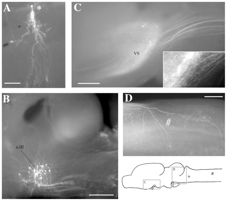
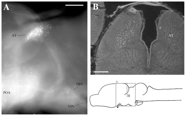
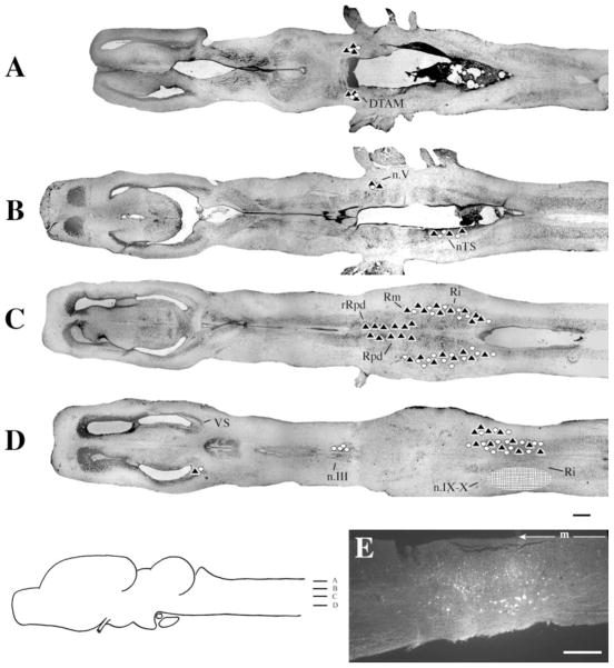
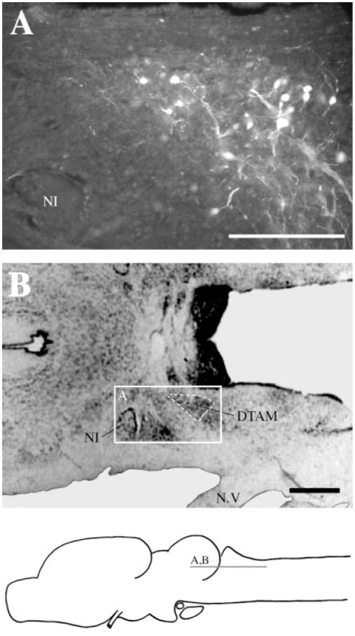
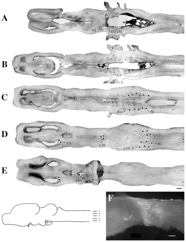
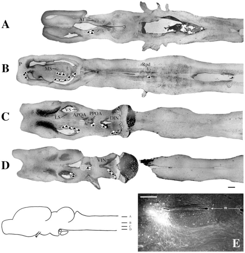
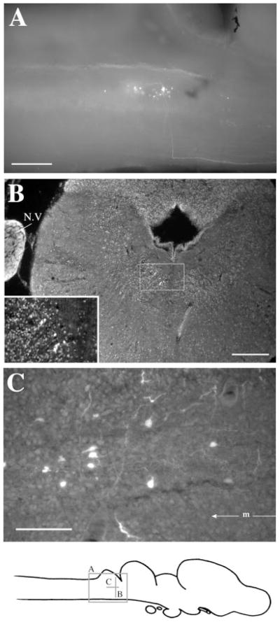
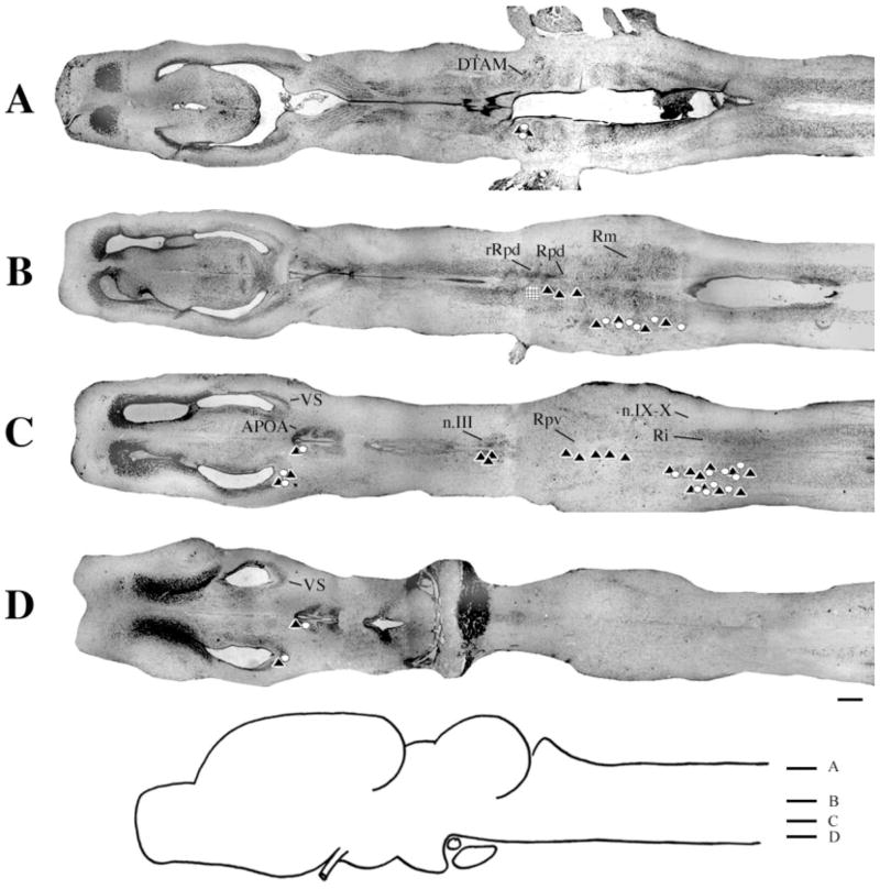
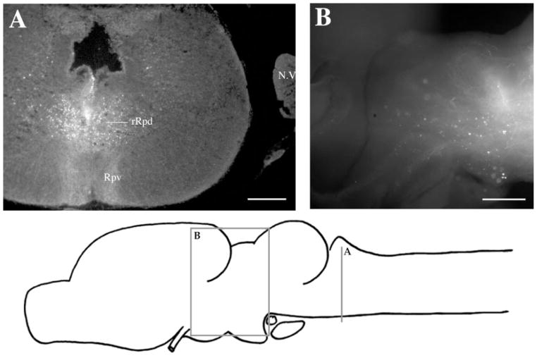
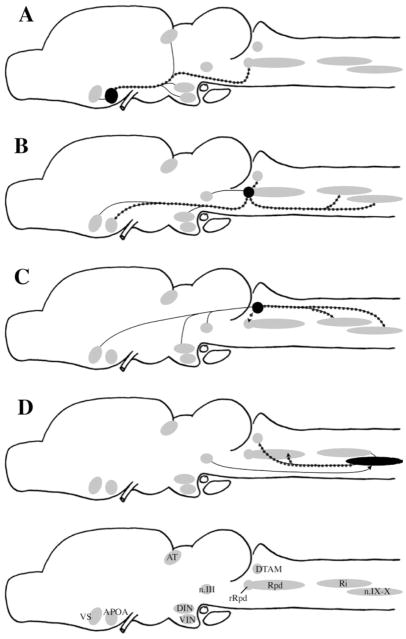
Sexually differentiated calling patterns of Xenopus laevis are conveyed to the vocal organ by a dedicated neuromuscular system. Here, we define afferents to vocal motor neurons and determine whether the connectivity of the vocal pathway is sexually differentiated. The use of fluorescent dextran amines and the isolated brain preparation readily permitted identification of anterograde and retrograde connectivity patterns. The whole-mount preparation allowed us to observe projections in their entirety, including cells of origin of a projection (for retrograde projections), terminal fields (for anterograde connections), and fiber tracts. Major findings are the confirmation of a robust and reciprocal connection between cranial nucleus (n.) IX-X and the pretrigeminal nucleus of the dorsal tegmental area of the medulla (DTAM) as well as between DTAM and the ventral striatum (VS). Newly revealed is the extensive connectivity between the rostral subdivision of the dorsal nucleus raphe (rRpd) and candidate vocal nuclei. In contrast to previous results using peroxidase, we did not observe dramatic sex differences in connectivity, although some connections were less robust in female than in male brains. Some retrograde connections previously observed (e.g., anterior preoptic area to DTAM) were not confirmed. Plausible hypotheses are that a set of rhombencephalic neurons located in DTAM, the inferior reticular formation and n.IX-X are responsible for generating patterned vocal activity, that activity is modulated by neurons in rRpd, and that activity in VS (particularly that evoked by conspecific calls), together with effects of steroid hormones at many sites in the vocal circuit, contribute to the initiation of calling.


Figures

Fig. 1
Fig. 1
Whole-mount preparations. A: A labeled cell in the Rpd after a discrete injection into rRpd. B: Labeled cell bodies in n.III after an injection into n.IX-X. Dendrites can be seen extending dorsoanteriorly and dorsoposteriorly. Posteriorly, labeled axons can be seen fasciculating ventrally before crossing to the contralateral side of the brain. C: Labeled terminal fields and cells in VS after an injection into DTAM. The projecting fiber tract can be seen posteriorly. Synaptic boutons and terminating fibers can be seen in the enlargement (insert). D: A labeled terminating fiber (#) in the medulla after an injection into DTAM. The locations of areas shown in C and B, the labeled cell in A, and the fiber in D are indicated on the sagittal diagram. For abbreviations, see list. Scale bars = 300 μm in A–D.
Fig. 2
Fig. 2
Label in AT after injecting APOA. A: Sagittal view in whole-mount showing labeled cells in AT, DIN, VIN, and POA as well as the projecting fiber tract to AT. B: Transverse section through AT. For abbreviations, see list. Scale bars = 300 μm in A,B.
Fig. 3
Fig. 3
Injections into n.IX-X. A–D: Black triangles indicate labeled terminal fields; white circles indicate labeled cells; the hatched area indicates the location and extent of a typical injection site. E: Photomicrograph of a typical injection site. m, midline. For other abbreviations, see list. Scale bar = 500 μm in D (applies to A–D), 300 μm in E.
Fig. 4
Fig. 4
Label in DTAM after Fluoro-Ruby injections into n.IX-X. A: Labeled cells, fibers, and synaptic boutons in a horizontal section. B: Reference cresyl-violet–stained horizontal section through DTAM. For abbreviation, see list. Scale bar = 300 μm in A, 500 μm in B.
Fig. 5
Fig. 5
Injections into DTAM. A–E: Symbols and scale bar as in Figure 3. F: Photomicrograph of a typical injection site. m, midline. For other abbreviations, see list. Scale bar = 300 μm in F.
Fig. 6
Fig. 6
Injections into APOA. A–D: Symbols and scale bar as in Figure 3. E: Photomicrograph of a typical injection site. m, midline. For other abbreviations, see list. Scale bar in E = 300 μm in E.
Fig. 7
Fig. 7
Label in rRpd after Fluoro-Ruby injections into APOA. A: Sagittal view in whole-mount. A labeled fiber can be seen traveling along the ventral surface from APOA and making a right-angled turn beneath the cerebellum and rRpd. B: Transverse section with labeled cells in rRpd. The insert is an enlargement of the boxed area and clearly shows seven labeled cells in rRpd. C: Labeled cells and fibers in a horizontal section through rRpd. m, midline. For other abbreviations, see list. Scale bars = 300 μm in A,B, 100 μm in C.
Fig. 8
Fig. 8
Injections into rRpd. Symbols and scale bar as in Figure 3. A typical injection site is illustrated in Figure 9A. For abbreviations, see list.
Fig. 9
Fig. 9
Label after Fluoro-Ruby injections into rRpd. A: Transverse section through the injection site, showing dense terminal fields in Rpv. B: Labeled cells and terminal fields in the thalamus in whole-mount. For abbreviations, see list. Scale bars = 300 μm in A,B.
Fig. 10
Fig. 10
Summary diagrams of results of injections into APOA (A), rRpd (B), DTAM (C), and n.IX-X (D). Plain lines indicate reciprocal connections. Dotted lines indicate projections that were confirmed by retrograde labeling. Arrows indicate direction of nonreciprocal projections. For abbreviations, see list.

References

    1. Allison JD, Wilczynski W. Thalamic and midbrain auditory projections to the preoptic area and ventral hypothalamus in the green treefrog (Hyla cinerea) Brain Behav Evol. 1991;38:322–331. - PubMed
    1. Beltz B, Eisen JS, Flamm R, Harris-Warrick RM, Hooper SL, Marder E. Serotonergic innervation and modulation of the stomatogastric ganglion of three decapod crustaceans (Panulirus interruptus, Homarus americanus, and Cancer irroratus) J Exp Biol. 1984;109:35–54. - PubMed
    1. Burmeister S, Wilczynski W. Social signals influence hormones independently of calling behavior in the treefrog (Hyla cinerea) Horm Behav. 2000;38:201–209. - PubMed
    1. Burmeister S, Somes C, Wilczynski W. Behavioral and hormonal effects of exogenous vasotocin and corticosterone in the green treefrog. Gen Comp Endocrinol. 2001;122:189–197. - PubMed
    1. Cardona A, Rudomin P. Activation of brainstem serotoninergic pathways decreases homosynaptic depression of monosynaptic responses of frog spinal motoneurons. Brain Res. 1983;280:373–378. - PubMed

Publication types

MeSH terms

LinkOut - more resources