

Review
doi: 10.1007/978-3-642-55580-0_8.
A novel vitamin D-regulated immediate-early gene, IEX-1, alters cellular growth and apoptosis
Affiliations
- PMID: 12899517
- PMCID: PMC2903742
- DOI: 10.1007/978-3-642-55580-0_8
Item in Clipboard
Review
A novel vitamin D-regulated immediate-early gene, IEX-1, alters cellular growth and apoptosis
Recent Results Cancer Res.
2003.
Abstract
1alpha,25-Dihydroxyvitamin D3 (1alpha,25(OH)2D3) inhibits the expression of an immediate-early gene, IEX-1, which is involved in the regulation of cellular growth and apoptosis in a variety of cells. 1alpha,25(OH)2D3 alters the subcellular localization of IEX-1 by causing an efflux of IEX-1 from the nucleus, and the sterol decreases the expression of IEX-1 messenger RNA in cells via a novel DR3 repeat-type DNA response element.
Figures
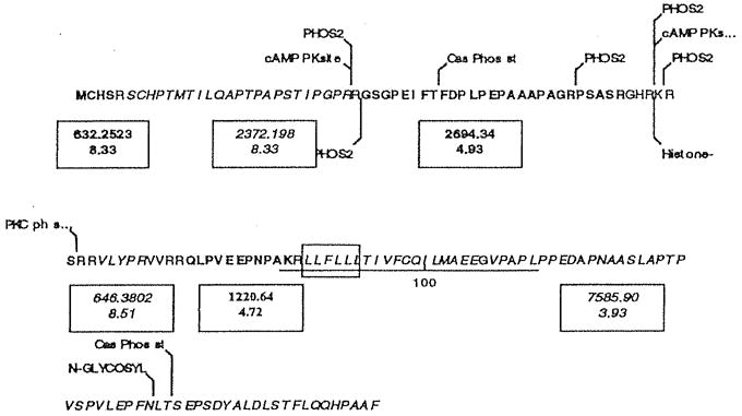
Amino acid sequence of IEX-1 (Swiss Prot Acc #P46695) and sites at which the protein might undergo posttranslational modification. The underlined sequence from residue 86 to 111 has homology with rhodopsin-like G protein coupled receptors, and the boxed amino acids from 88 to 93 display the LLXLL motif characteristic of coactivator proteins. A nuclear localization signal, RKRSRR partially overlaps the PKC phosphorylation site. Also shown, in alternating bold and italics, are the tryptic fragments greater than 500 Da, with the monoistopic mass and pl of each unmodified fragment. These fragments represent approximately 93% of the 156-amino acid protein. Calculated molecular weight, 16,927.48 kDa; estimated pl, 8.83
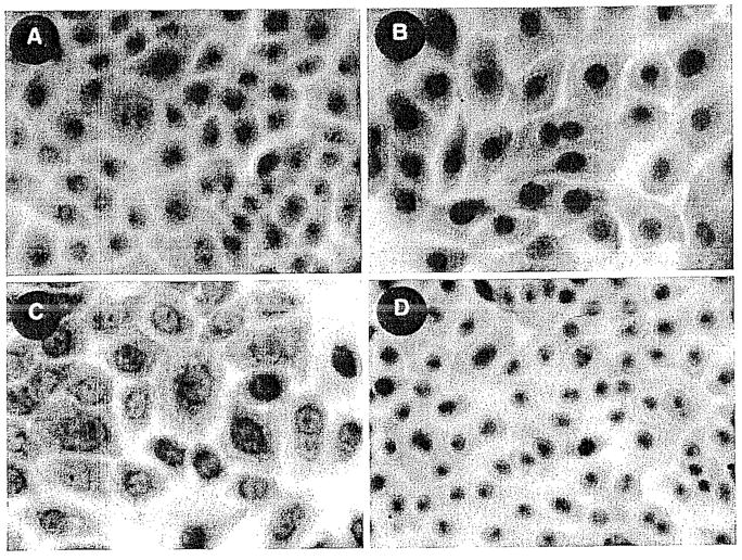
A–D. IEX-1 Immunostaining in normal keratinocytes. A IEX-1 localized within nucleus. B Negative control. C IEX-1 localized in nuclear and perinuclear regions. D Negative control. All cells were fixed with methanol-acetone.
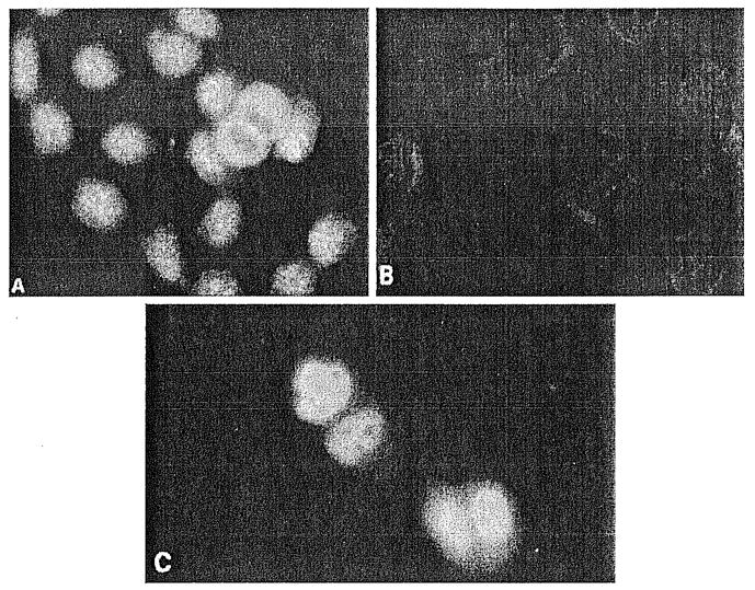
A–C Fluorescent microscopy of HaCaT cells localizing IEX-1 protein. Replicating HaCaT cells stained with: A IEX-1-1-specific antibody; B preimmune serum; C HaCaT cells transfected with GFP-IEX-1 chimeric plasmid and visualized directly by fluorescent microscopy 48 h later. (From Kumar et al. 1998, with permission)
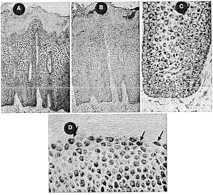
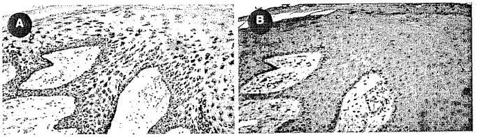
A–D IEX-1 immunostaining in psoriatic skin. A Hyperplastic and acanthotic epidermis showing intense IEX-1 staining within basal cell and suprabasal layers. B Negative control, no primary Ab. C High-power rete peg showing strong perinuclear staining. D Upper granular layer with strong nuclear localization of IEX-1 protein.
A, B IEX-1 Immunostaining in squamous cell carcinoma. A IEX-1 staining within hyperplastic tumor fronds. B Negative control.
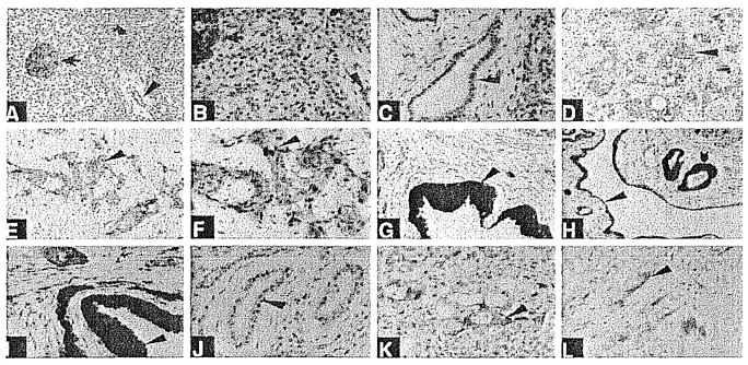
A–L. Immunohistochemistry of pancreatic tissues using a specific antigen-affinity purified antibody to IEX-1 (A–I, K). Immunostaining in J and L is with IEX-1 antibody preabsorbed with antigen. A normal pancreatic tissue (×200). Arrow shows intense staining of an islet of Langerhans. Arrowhead shows modest staining of ductal epithelium. Blocked arrow shows some staining of acinar cells. B Normal pancreatic tissue (×400). Arrows indicate same tissue as in A. C Normal pancreatic tissue (×400). Note immunostaining of some ductal epithelial cells. D, E Pancreatic acinar adenocarcinoma (×200). Note immunostaining of tumor cells (arrowhead). F Pancreatic acinar adenocarcinoma (×400). Note immunostaining of tumor cells (arrowhead). G Chronic pancreatitis (×200). Note immunostaining of hyperplastic ductal epithelial cells (arrowhead). Nuclear and cytoplasmic staining is evident. H Chronic pancreatitis (×200). Note immunostaining of hyperplastic ductal-epitheliai cells (blocked arrow and arrowhead). Nuclear and cytoplasmic staining is evident. Not all ductal cells are as intensely stained. I Chronic pancreatitis (×400). Note immunostaining of hyperplastic ductal epithelial cells (arrowhead). Nuclear and cytoplasmic staining is evident. Not all ductal cells are as intensely stained. J Chronic pancreatitis (×200). Preabsorbed serum. K Pancreatic carcinoma metastatic to the liver (×400). Note immunostaining of carcinoma cells (arrowhead). L Pancreatic acinar adenocarcinoma (×400). Preadsorbed antiserum.
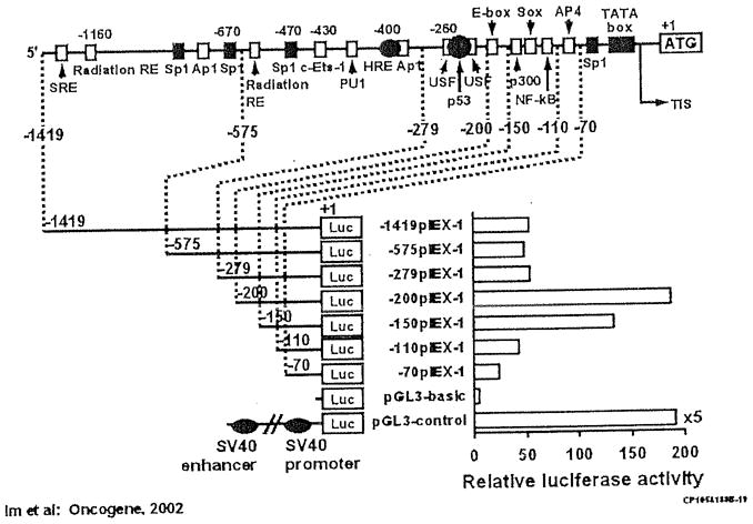
Positions of putative transcription factor binding sites discussed in the text are indicated. Putative HRE (VDRE) is shown as black oval. A TATA box is located 25 bp upstream from the transcription initiation site (TIS). Promoter activities of the IEX-1 promoter deletion constructs fused to firefly luciferase reporter gene transiently transfected in HaCaT cells. Left, the length of the tested promoter fragments is shown. Numbers indicate the relative positions with respect to the transcription start site. Right, luciferase activity is shown as n-fold value compared to cells transfected with the promoterless pGL3-basic vector (which was assigned an activity value of 1.0). Data represent the means of three independent experiments in duplicates, with at least two different plasmid preparations
Similar articles
-
Characterization of a novel hexameric repeat DNA sequence in the promoter of the immediate early gene, IEX-1, that mediates 1alpha,25-dihydroxyvitamin D(3)-associated IEX-1 gene repression.Oncogene. 2002 May 23;21(23):3706-14. doi: 10.1038/sj.onc.1205450. Oncogene. 2002. PMID: 12032839
-
Regulation of a novel immediate early response gene, IEX-1, in keratinocytes by 1alpha,25-dihydroxyvitamin D3.Biochem Biophys Res Commun. 1998 Oct 29;251(3):868-73. doi: 10.1006/bbrc.1998.9556. Biochem Biophys Res Commun. 1998. PMID: 9791001
-
Immediate early gene X1 (IEX-1) is organized in subnuclear structures and partially co-localizes with promyelocytic leukemia protein in HeLa cells.J Biol Chem. 2005 Jul 1;280(26):24849-56. doi: 10.1074/jbc.M501571200. Epub 2005 Apr 26. J Biol Chem. 2005. PMID: 15855159
-
Roles of the stress-induced gene IEX-1 in regulation of cell death and oncogenesis.Apoptosis. 2003 Jan;8(1):11-8. doi: 10.1023/a:1021688600370. Apoptosis. 2003. PMID: 12510147 Review.
-
Regulation of primary response genes.Mol Cell. 2011 Nov 4;44(3):348-60. doi: 10.1016/j.molcel.2011.09.014. Mol Cell. 2011. PMID: 22055182 Free PMC article. Review.
Cited by
-
HCMV miRNA Targets Reveal Important Cellular Pathways for Viral Replication, Latency, and Reactivation.Noncoding RNA. 2018 Oct 22;4(4):29. doi: 10.3390/ncrna4040029. Noncoding RNA. 2018. PMID: 30360396 Free PMC article. Review.
-
The role of Iex-1 in the pathogenesis of venous neointimal hyperplasia associated with hemodialysis arteriovenous fistula.PLoS One. 2014 Jul 18;9(7):e102542. doi: 10.1371/journal.pone.0102542. eCollection 2014. PLoS One. 2014. PMID: 25036043 Free PMC article.
-
Immediate early gene X-1 interacts with proteins that modulate apoptosis.Biochem Biophys Res Commun. 2004 Oct 29;323(4):1293-8. doi: 10.1016/j.bbrc.2004.09.006. Biochem Biophys Res Commun. 2004. PMID: 15451437 Free PMC article.
-
Immediate early response gene X-1, a potential prognostic biomarker in cancers.Expert Opin Ther Targets. 2013 May;17(5):593-606. doi: 10.1517/14728222.2013.768234. Epub 2013 Feb 4. Expert Opin Ther Targets. 2013. PMID: 23379921 Free PMC article. Review.
-
Rearrangements and amplification of IER3 (IEX-1) represent a novel and recurrent molecular abnormality in myelodysplastic syndromes.Cancer Res. 2009 Oct 1;69(19):7518-23. doi: 10.1158/0008-5472.CAN-09-1428. Epub 2009 Sep 22. Cancer Res. 2009. PMID: 19773435 Free PMC article.
References
-
- Arlt A, Grobe O, Sieke A, Kruse ML, Folsch UR, Schmidt WE, Schafer H. Expression of the NF-kappa B target gene IEX-1 (p22/PRG1) does not prevent cell death but instead triggers apoptosis in Hela cells. Oncogene. 2001;20:69–76. - PubMed
-
- Beckman MJ, DeLuca HF. Modern view of vitamin D3 and its medicinal uses. Prog Med Chem. 1998;35:1–56. - PubMed
-
- Bellido T, Boland R. Stimulation of myoblast membrane protein synthesis by 25-hydroxy-vitamin D3. Zeitschrift fur Naturforschung. Section C. J Biosci. 1989;44:807–812. - PubMed
-
- Bellido T, Morelli S, Fernandez LM, Boland R. Evidence for the participation of protein kinase C and 3′,5′-cyclic AMP-dependent protein kinase in the stimulation of muscle cell proliferation by 1,25-dihydroxy-vitamin D3. MolCell Endocrinol. 1993;90:231–28. - PubMed
-
- Bikle DD. Clinical counterpoint: vitamin D: new actions, new analogs, new therapeutic potential. Endocr Rev. 1992;13:765–784. - PubMed
Publication types
MeSH terms
Substances
Grants and funding
LinkOut - more resources
Full Text Sources