

Review
doi: 10.1016/s0952-7915(03)00079-7.
Real-time imaging of lymphocytes in vivo
Affiliations
- PMID: 12900266
- PMCID: PMC2953535
- DOI: 10.1016/s0952-7915(03)00079-7
Item in Clipboard
Review
Real-time imaging of lymphocytes in vivo
Curr Opin Immunol.
2003 Aug.
Abstract
New preparations, fluorescent probes and imaging techniques are providing the means to observe the behavior of cells in the tissue environment of lymphoid organs. In particular, when combined with two-photon laser microscopy, intravital imaging of surgically exposed lymph nodes provides a unique view of lymphocyte migration and antigen presentation as it occurs within the living animal. The view is emerging that lymphocytes migrate randomly within lymphoid organs, and that lymphocyte contact with antigen-presenting cells may be a stochastic process rather than one guided by chemokine gradients.
Figures
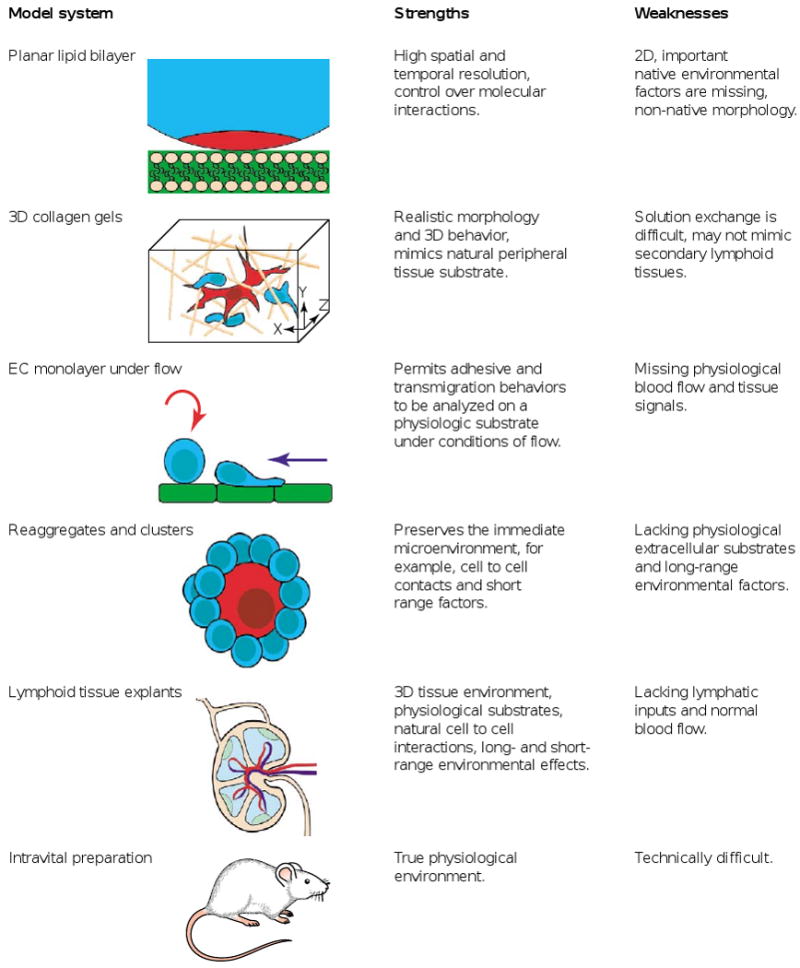
The strengths and weaknesses of model systems in current use for the real-time imaging of lymphocytes. EC, endothelial cell.
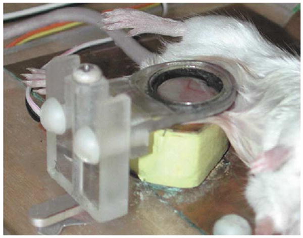
Intravital two-photon microscopy: an anesthetized mouse with surgically exposed lymph node on the microscope stage. Details of the preparation have been described [23].
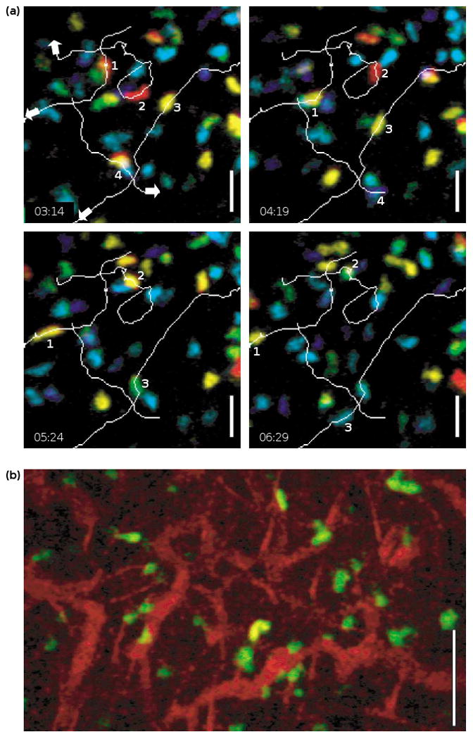
Intravital two-photon images of T cells in the inguinal lymph node of an anesthetized mouse. (a) Trajectories of four separate cells at varying times. The colors represent cells at different depths, ranging from ∼100 to 150 μm below the surface of the lymph node, with blue representing the bottom and red the top of the imaging volume. Scale bar: 25 μm. (b) T cells (green), vessels and fibers (both red) labeled via tail vein injection with tetramethylrhodamine dextran. Scale bar: 50 μm.
References
-
- Gretz JE, Anderson AO, Shaw S. Cords, channels, corridors and conduits: critical architectural elements facilitating cell interactions in the lymph node cortex. Immunol Rev. 1997;156:11–24. - PubMed
-
- Gretz JE, Norbury CC, Anderson AO, Proudfoot AE, Shaw S. Lymph-borne chemokines and other low molecular weight molecules reach high endothelial venules via specialized conduits while a functional barrier limits access to the lymphocyte microenvironments in lymph node cortex. J Exp Med. 2000;192:1425–1440. - PMC - PubMed
-
- Cyster JG. Chemokines and cell migration in secondary lymphoid organs. Science. 1999;286:2098–2102. - PubMed
-
- Jenkins MK, Khoruts A, Ingulli E, Mueller DL, McSorley SJ, Reinhardt RL, Itano A, Paper KA. In vivo activation of antigen-specific CD4 T cells. Annu Rev Immunol. 2001;19:23–45. - PubMed
-
- Grakoui A, Bromley SK, Sumen C, Davis MM, Shaw AS, Allen PM, Dustin ML. The immunological synapse: a molecular machine controlling T cell activation. Science. 1999;285:221–227. - PubMed
Publication types
MeSH terms
Grants and funding
LinkOut - more resources
Full Text Sources
Other Literature Sources
