. 2003 Aug 4;162(3):383-90.
doi: 10.1083/jcb.200301073.

Microtubules are the only structural constituent of the spindle apparatus required for induction of cell cleavage

Affiliations

Microtubules are the only structural constituent of the spindle apparatus required for induction of cell cleavage

G Bradley Alsop et al. J Cell Biol. .

Abstract

Structural constituents of the spindle apparatus essential for cleavage induction remain undefined. Findings from various cell types using different approaches suggest the importance of all structural constituents, including asters, the central spindle, and chromosomes. In this study, we systematically dissected the role of each constituent in cleavage induction in grasshopper spermatocytes and narrowed the essential one down to bundled microtubules. Using micromanipulation, we produced "cells" containing only asters, a truncated central spindle lacking both asters and chromosomes, or microtubules alone. We show that furrow induction occurs under all circumstances, so long as sufficient microtubules are present. Microtubules, as the only spindle structural constituent, undergo dramatic, stage-specific reorganizations, radiating toward cell cortex in "metaphase," disassembling in "anaphase," and bundling into arrays in "telophase." Furrow induction usually occurs at multisites around microtubule bundles, but only those induced by sustained bundles ingress. We suggest that microtubules, regardless of source, are the only structural constituent of the spindle apparatus essential for cleavage furrow induction.


Figures

Figure 1.
Figure 1.
Asters alone are sufficient for induction of cell cleavage. (A) Asters are detached from spindle poles using a microneedle (a), placed at the cell periphery (b), and secluded into a membrane pocket cut off from the mother cell (c and d). (B) Polarization microscope images. Time is given in minutes. The membrane pocket contains two prominent asters (0 min, *) with mitochondria on one side (m). The mother cell (left) bears the spindle with truncated poles. Formation of microtubule– mitochondria bundles in the pocket (31), similar to the central spindle in its mother cell, corresponds with disappearance of asters. Furrow initiation of the pocket and mother cell occurs simultaneously (63). The pocket initiates a furrow where the bundled microtubule array is proximal to the cortex (63, arrow), ingressing asymmetrically on one side of the cell (93, arrow). (C) Symmetric furrow ingression can also occur in an aster-containing membrane pocket (0–62; see Video 1, available at http://www.jcb.org/cgi/content/full/jcb.200301073/DC1). (D) Microtubules (MT) at furrow initiation are less organized than observed in spindle-containing cells, but do exhibit a lightly stained midzone (MT, arrow) enriched with actin filaments (AF and Overlay, arrow). (E) Once ingressed, the bundled microtubule array establishes a clear midzone (MT, arrows) embraced with a tightly organized contractile ring (AF and Overlay). (F) Disassembly of astral microtubules (0, *) with nocodazole treatment (11 onward) prevents furrow induction in an aster-containing pocket (11–74) that bears only a disorganized mitochondrial mass. Although random contractions can still occur (74 onward), no furrow initiation is observed. Bars, 10 μm.
Figure 2.
Figure 2.
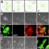
Truncated central spindles alone are sufficient for cell cleavage. (A) Spindle poles, including asters and pronuclei, are removed from late anaphase or early telophase cells (a–c). The remaining central spindle fragment is then rotated ∼90° from the original equator to avoid any predeposited furrow signals (d). (B) A truncated central spindle (0 min onward), lacking astral microtubules (MT) and chromosomes (DAPI), initiates a furrow (10 min, arrows) around the spindle midzone (MT, arrows) enriched with actin filaments (AF and Overlay, arrows). (C) Furrow ingression (0–16) is normal, bearing a distinct contractile ring at the midzone of the truncated spindle (Overlay). Bars, 10 μm.
Figure 3.
Figure 3.
Microtubules alone are sufficient for cell cleavage. (A) Asters and chromosomes are removed in metaphase (a–c), which induces disassembly of the spindle (d). (B) After manipulation, the spindle collapses (0–37 min; see Video 2, available at http://www.jcb.org/cgi/content/full/jcb.200301073/DC1), inducing transient and repeated formations of bipolar (70 and 194) or monopolar (217) pseudospindles, and eventually gives rise to relatively disorganized arrays of bundled microtubules bearing pseudopoles (290–296, *). Furrow initiation (290–296, arrows) and accumulation of actin filaments (AF and Overlay) correspond to the midzone (MT, arrows) of bundled microtubule arrays. Due to microtubule reorganizations, furrow initiation is delayed by nearly 2 h in comparison with an initially synchronized cell (183, arrow). (C) Often, newly assembled microtubule bundles radiate randomly (0–108) and induce furrow initiation at multiple sites (118–128, arrows and arrowheads; see Video 3, available at http://www.jcb.org/cgi/content/full/jcb.200301073/DC1). However, only furrows initiated by persistent bundles sustain (118–137, arrows), others eventually regress (arrowheads). Furrow ingression forces the bundled microtubule arrays together (128–153), bearing a single midzone (MT, arrow) with an ingressed contractile ring (AF and Overlay). Furrow initiation is delayed by ∼1 h, judging by division in a neighboring cell (43, arrow). Bar, 10 μm.
Figure 4.
Figure 4.
Bundled microtubule arrays are required for furrow induction. Cells were manipulated as in Fig. 3 to remove both asters and chromosomes, inducing assembly of bundled microtubules. (A) Partial disassembly of microtubules with nocodazole at furrow initiation (88 min) leads to regression of the furrow (101 onward). The fixed cell (116) retains remnants of bundled microtubule arrays (MT) and actin filaments from the disassembled contractile ring (AF and Overlay, arrow). (B) Complete disassembly of microtubules (MT) with nocodazole before furrow initiation (17) results in formation of a disorganized mitochondrial mass (44 onward). Although random contractions can still occur (101–187), no furrow initiation is observed. Actin filaments (AF and Overlay) are associated primarily with the mitochondrial mass. Bar, 10 μm.
Figure 5.
Figure 5.
Furrow abscission and regression in micromanipulated cells. Unless indicated, time is given in minutes. (A) An aster-containing pocket, produced as in Fig. 1, undergoes successful furrow initiation (0–68 min, arrows) and ingression (88), but fails in abscission due to furrow regression (16 h). (B) A cell lacking both asters and pronuclei, manipulated as in Fig. 2, is fully capable of furrow induction (12–45, arrows), ingression (72), and normal abscission (17.1 h; see Video 4, available at http://www.jcb.org/cgi/content/full/jcb.200301073/DC1). (B') Rhodamine dextran microinjected into one daughter cell gradually flows into the other in both visually abscised experimental cells (aster removal, 17.3–17.8 h after cleavage initiation) and nonmanipulated controls (control, 17.2–17.7 h after cleavage initiation). (C) Despite normal furrow induction and ingression (155–243, arrows), cells containing only microtubules, manipulated as in Fig. 3, usually fail to separate (315–390, arrow; see Video 5, available at http://www.jcb.org/cgi/content/full/jcb.200301073/DC1). An exception is shown in Fig. S2, available at http://www.jcb.org/cgi/content/full/jcb.200301073/DC1. Bars, 10 μm.

References

    1. Bonaccorsi, S., M.G. Giansanti, and M. Gatti. 1998. Spindle self-organization and cytokinesis during male meiosis in asterless mutants of Drosophila melanogaster. J. Cell Biol. 142:751–761. - PMC - PubMed
    1. Bucciarelli, E., M.G. Giansanti, S. Bonaccorsi, and M. Gatti. 2003. Spindle assembly and cytokinesis in the absence of chromosomes during Drosophila male meiosis. J. Cell Biol. 160:993–999. - PMC - PubMed
    1. Bukauskas, F.F., C. Kempf, and R. Weingart. 1992. Cytoplasmic bridges and gap junctions in an insect cell line (Aedes albopictus). Exp. Physiol. 77:903–911. - PubMed
    1. Canman, J.C., D.B. Hoffman, and E.D. Salmon. 2000. The role of pre- and post-anaphase microtubules in the cytokinesis phase of the cell cycle. Curr. Biol. 10:611–614. - PubMed
    1. Cao, L., and Y.L. Wang. 1996. Signals from the spindle midzone are required for the stimulation of cytokinesis in cultured epithelial cells. Mol. Biol. Cell. 7:225–232. - PMC - PubMed

Publication types