. 2003 Aug 19;100(17):10108-13.
doi: 10.1073/pnas.1733208100. Epub 2003 Aug 4.

The VirD2 pilot protein of Agrobacterium-transferred DNA interacts with the TATA box-binding protein and a nuclear protein kinase in plants

Affiliations

The VirD2 pilot protein of Agrobacterium-transferred DNA interacts with the TATA box-binding protein and a nuclear protein kinase in plants

László Bakó et al. Proc Natl Acad Sci U S A. .

Abstract

The bacterial virulence protein VirD2 plays an important role in nuclear import and chromosomal integration of Agrobacterium-transferred DNA in fungal, plant, animal, and human cells. Here we show that in nuclei of alfalfa cells, VirD2 interacts with and is phosphorylated by CAK2Ms, a conserved plant ortholog of cyclin-dependent kinase-activating kinases. CAK2Ms binds to and phosphorylates the C-terminal regulatory domain of RNA polymerase II largest subunit, which can recruit the TATA box-binding protein. VirD2 is found in tight association with the TATA box-binding protein in vivo. These results indicate that recognition of VirD2 is mediated by widely conserved nuclear factors in eukaryotes.


Figures
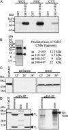
Fig. 1.
Fig. 1.
VirD2 is phosphorylated by and interacts with a nuclear protein kinase. (A) In vitro protein kinase assays with alfalfa whole-cell extract (WCE) and purified nuclear (NUC) and cytoplasmic (CYT) protein fractions by using GST and GST-VirD2 substrates. (B) Cyanbromide fragmentation of GST-VirD2 after phosphorylation with alfalfa nuclear extract shows 32P labeling of peptides located between VirD2 amino acid positions 2–109 (12.2 kDa) and 248–447 (22 kDa). (C) Expression of HA-VirD2 detected by Western blotting with anti-HA IgG in protoplasts 12, 24, and 36 h after transformation with pRT100-HA-VirD2 sense DNA. Controls to the left show HA-VirD2 purified from E. coli and a lack of HA-VirD2 expression in cells transformed with an antisense construct. (D) Immunoprecipitation of 32P-labeled HA-VirD2 from alfalfa protoplasts. After Coomassie blue staining (Left), the gel was subjected to autoradiography (Right). [32P]HA-VirD2 shows similar mobility as HA-VirD2 purified from E. coli. VH and VL are the heavy and light chains of IgG.
Fig. 2.
Fig. 2.
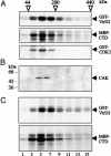
Nuclear VirD2-kinase fractions purified by glycerol-gradient centrifugation phosphorylate RNA polymerase II CTD and CDK2 and are immunoprecipitated by anti-R2Os IgG. (A) Phosphorylation assays with glycerol-gradient fractions of nuclear VirD2-kinase using GST-VirD2, MBP-CTD, and GST-CDK2 substrates in vitro. (B) Western blotting of glycerol-gradient fractions with anti-R2Os IgG. (C) From the glycerol-gradient fractions, anti-R2Os IgG immunoprecipitates a protein kinase that phosphorylates GST-VirD2 and GST-CTD in vitro. Positions of glycerol-gradient and SDS/PAGE size markers (kDa) are shown above A and to the left of B.
Fig. 3.
Fig. 3.
CAK2Ms, a close homolog of Arabidopsis CAK2At and rice R2Os protein kinases, interacts with and phosphorylates VirD2 and RNA polymerase II CTD. (A) Alignment of CAK2Ms (AAK97227), R2Os (CAA41172), and CAK2At (BAB62843) kinase sequences. Frames mark regions with identical amino acids. (B)(Left) Detection of HA-CAK2Ms expression in alfalfa protoplasts by immunoblotting with anti-HA IgG after transformation with pPE1000-HA-CAK2Ms DNA. Cells transformed with the empty vector pPE1000 show no signal. (Right) Immunoprecipitation of HA-CAK2Ms from alfalfa protein extracts in the presence (+) or absence (–) of competitor HA-peptide, followed by kinase assays with GST-VirD2. (C) VirD2-kinase from the glycerol-gradient fractions (Fig. 2) is selectively retained on GST-VirD2 (Left) and MBP-CTD (Right) affinity resins and phosphorylates these substrate proteins in kinase pull-down assays.
Fig. 4.
Fig. 4.
Molecular interactions among VirD2, TBP, and RNA polymerase II CTD. (A) SDS/PAGE analysis of in vitro protein interactions shows that MBP-CTD binds TBP, but not VirD2, whereas VirD2 recruits TBP. (B)(Upper) His6-TBP forms a salt-resistant complex with GST-VirD2 but does not bind specifically to control glutathione-Sepharose and GST-loaded beads. (Lower) The N-terminal VirD2 domain is necessary for TBP binding. GST-VirD2ΔNT, carrying an N-terminal VirD2 deletion of 266 aa, and the control GST protein show only trace levels of TBP binding. GST-VirD2 and GST-VirD2ΔCT, carrying a C-terminal VirD2 deletion, show strong interaction with TBP. (C) Transient expression of HA-VirD2 by Agrobacterium transformation in Arabidopsis cells. Multiple forms of HA-VirD2 protein are observed by immunoblotting with anti-HA and anti-VirD2 IgGs, as well as by anti-HA immunoprecipitation (αHA-IP), followed by Western blotting with anti-VirD2 IgG. (D) (Left) Detection of TBP in wild-type cells by Western blotting with anti-TBP IgG. (Right) Detection of TPB after immunoprecipitation of protein extracts from wild-type cells (wt) and HA-VirD2-expressing Agrobacterium-transformed Arabidopsis cells with anti-TBP Ab.

References

    1. Gelvin, S. B. (2000) Annu. Rev. Plant Physiol. Plant Mol. Biol. 51, 223–256. - PubMed
    1. Tzfira, T., Rhee, Y., Chen, M.-H., Kunik, T. & Citovsky, V. (2000) Annu. Rev. Microbiol. 54, 187–219. - PubMed
    1. Zambryski, P., Tempé, J. & Schell, J. (1989) Cell 56, 193–201. - PubMed
    1. Kitakura, S., Fujita, T., Ueno, Y., Terakura, S., Wabiko, H. & Machida, Y. (2002) Plant Cell 14, 451–463. - PMC - PubMed
    1. Bundock, P., den Dulk-Das, A., Beijersbergen, A. & Hooykaas, P. J. J. (1995) EMBO J. 14, 3206–3214. - PMC - PubMed

Publication types

MeSH terms

Associated data