. 2003 Aug 4;198(3):483-9.
doi: 10.1084/jem.20022027.

VEGF164-mediated inflammation is required for pathological, but not physiological, ischemia-induced retinal neovascularization

Affiliations

VEGF164-mediated inflammation is required for pathological, but not physiological, ischemia-induced retinal neovascularization

Susumu Ishida et al. J Exp Med. .

Abstract

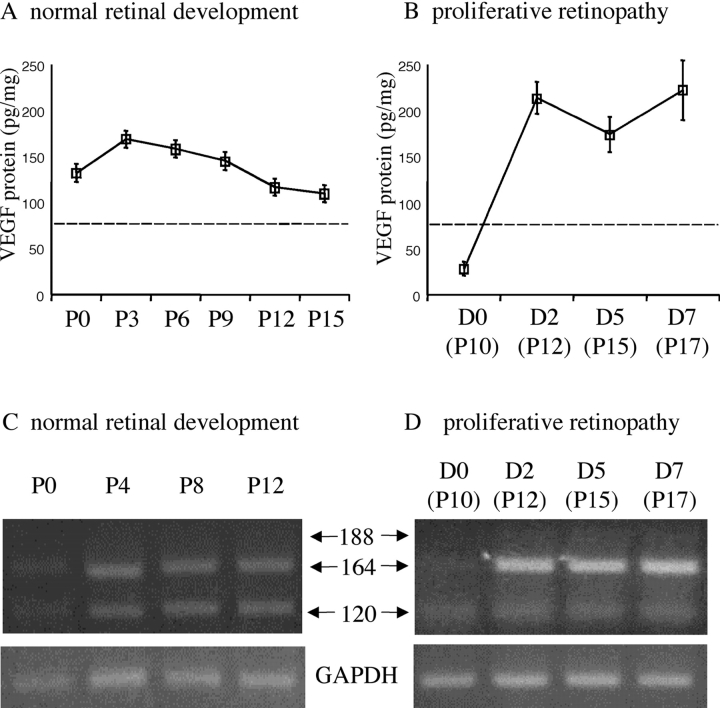
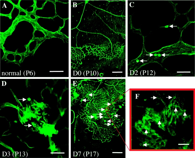
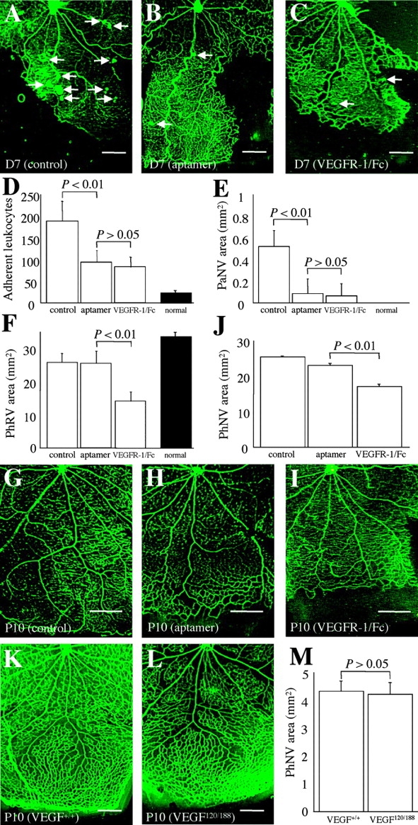
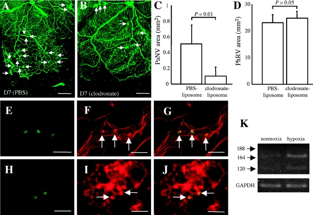
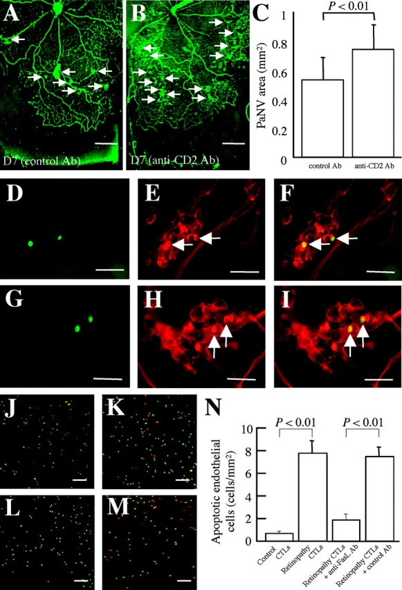
Hypoxia-induced VEGF governs both physiological retinal vascular development and pathological retinal neovascularization. In the current paper, the mechanisms of physiological and pathological neovascularization are compared and contrasted. During pathological neovascularization, both the absolute and relative expression levels for VEGF164 increased to a greater degree than during physiological neovascularization. Furthermore, extensive leukocyte adhesion was observed at the leading edge of pathological, but not physiological, neovascularization. When a VEGF164-specific neutralizing aptamer was administered, it potently suppressed the leukocyte adhesion and pathological neovascularization, whereas it had little or no effect on physiological neovascularization. In parallel experiments, genetically altered VEGF164-deficient (VEGF120/188) mice exhibited no difference in physiological neovascularization when compared with wild-type (VEGF+/+) controls. In contrast, administration of a VEGFR-1/Fc fusion protein, which blocks all VEGF isoforms, led to significant suppression of both pathological and physiological neovascularization. In addition, the targeted inactivation of monocyte lineage cells with clodronate-liposomes led to the suppression of pathological neovascularization. Conversely, the blockade of T lymphocyte-mediated immune responses with an anti-CD2 antibody exacerbated pathological neovascularization. These data highlight important molecular and cellular differences between physiological and pathological retinal neovascularization. During pathological neovascularization, VEGF164 selectively induces inflammation and cellular immunity. These processes provide positive and negative angiogenic regulation, respectively. Together, new therapeutic approaches for selectively targeting pathological, but not physiological, retinal neovascularization are outlined.


Figures

Figure 1.
Figure 1.
Differential expression of VEGF isoforms during physiological and pathological retinal neovascularization. (A) Retinal VEGF protein levels (all isoforms) during postnatal development. (B) Retinal VEGF protein levels after the induction (D0) of pathological neovascularization. Dotted lines in A and B indicate the constitutive VEGF levels in normal adult rats. (C) Retinal VEGF mRNA expression during postnatal development. (D) Retinal VEGF mRNA expression after the induction of pathological neovascularization. The relative expression levels of VEGF164 increased dramatically during pathological neovascularization.
Figure 2.
Figure 2.
Leukocytes are present at the leading edge of pathological, but not physiological, retinal neovascularization. (A) No leukocyte adhesion at the leading edge of physiological retinal development on P6. (B–F) Leukocyte adhesion during the onset and progression of pathological neovascularization. (B) The formation of extensive retinal ischemia on D0, immediately after exposure to relative hypoxia (room air). (C) Abundant inflammatory cells (arrows) randomly adhered to the intraretinal vessels just before the onset of pathological neovascularization. (D) The leukocyte adhesion (arrows) associated with the incipient vascular budding on D3. (E) Compared with D0 (B), revascularization proceeded in concert with pathological neovascularization (arrows) on D7. (F) Leukocyte adhesion (arrows) spatially and temporally associated with vascular budding. Bars: (A, C, D, and F) 50 μm; (B and E) 0.5 mm.
Figure 3.
Figure 3.
Effect of VEGF164-specific blockade on physiological and pathological retinal neovascularization. (A–F) VEGF164-specific blockade versus VEGF pan-isoform blockade in pathological neovascularization. (A) Pathological neovascularization (arrows, D7) treated with PEG control (n = 12) was not inhibited. (B) Pathological neovascularization (arrows, D7) treated with the anti-VEGF165 aptamer (n = 13). (C) Pathological neovascularization (arrows, D7) treated with the VEGFR-1/Fc chimera (n = 10). In addition to the inhibition of leukocyte adhesion to the retinal vasculature (D, P < 0.01), pathological neovascular budding into the vitreous (E, PaNV) was significantly suppressed (P < 0.01) via the anti-VEGF164 aptamer or the VEGFR-1/Fc. In contrast, the effect of VEGF164 inhibition on physiological revascularization (F, PhRV) was negligible (P > 0.05), but pan-isoform inhibition led to significant suppression of revascularization (F, P < 0.01). Shaded bars indicate comparable values of age-matched (P17) normal rat neonates (n = 8). (G–J) VEGF164-specific blockade versus VEGF pan-isoform blockade in retinal vascular development. (G) Developing retinal vasculature (P10) treated with PEG control (n = 7). (H) Developing retinal vasculature (P10) treated with anti-VEGF165 aptamer (n = 8). (I) Developing retinal vasculature (P10) treated with the VEGFR-1/Fc chimera (n = 10). (J) Note the mild suppression of physiological neovascularization during retinal development via VEGF164 inhibition, but the substantial suppression via pan-isoform inhibition (P < 0.01). (K–M) VEGF164-specific deficiency versus wild type in retinal vascular development. (K) Developing retinal vasculature (P10) in wild-type control (VEGF+/+) mice (n = 13). (L) Developing retinal vasculature (P10) in VEGF164-deficient (VEGF120/188) mice (n = 13). (M) VEGF164 deficiency had no significant effect on physiological neovascularization (P > 0.05). Bars: (K and L) 0.2 mm; (A–C and G–I) 0.5 mm.
Figure 4.
Figure 4.
Role of monocytes in pathological retinal neovascularization. (A) Pathological neovascularization (arrows, D7) treated with PBS control-liposomes (n = 8) was not inhibited. (B) Pathological neovascularization (arrows, D7) treated with clodronate-liposomes (n = 8). Notably, the pathological neovascular budding (C, PaNV) was suppressed (P < 0.01), whereas revascularization (D, phRV) was not (P > 0.05). (E–J) Monocyte adhesion was observed just before and during pathological neovascularization (H–J). Green fluorescence from the anti-CD13 antibody (E and H) and red fluorescence from the rhodamine-coupled Con A (F and I) identifies the Con A–stained cells as being CD13-positive leukocytes (arrows) when the images were superimposed (G and J). (K) Monocyte VEGF mRNA expression in normoxia (21% oxygen) and hypoxia (1% oxygen). VEGF levels were markedly increased in response to hypoxic stimulation. Bars: (E–J) 50 μm; (A and B) 0.5 mm.
Figure 5.
Figure 5.
Role of T lymphocytes in pathological retinal neovascularization. (A) Pathological neovascularization (arrows, D7) treated with nonimmune isotype control (n = 9) showing a similar degree of pathological vascular budding (arrows) compared with Fig. 2 E. (B) Pathological neovascularization (arrows, D7) treated with anti-CD2 antibody (n = 11). Notably, the pathological neovascular budding (C, PaNV) was worsened (P < 0.01). (D–I) T cell subtypes in pathological neovascular buds. Green fluorescence from the antibody against CD8 or CD25 (D and G) and red fluorescence from the rhodamine-coupled Con A (E and H) identifies the Con A–stained cells as being CD8- and CD25-positive leukocytes (arrows) when the images were superimposed (F and I). (J–N) Rhodamine-labeled apoptotic cells in the leukocyte–endothelial cocultures were detected via TUNEL staining (red). CTLs were labeled with CFDASE (green). Compared with control CTLs (J), retinopathy CTLs (K) significantly increased the number of TUNEL-positive endothelial cells (N, P < 0.01). The process was significantly inhibited via FasL blockade (L–N, n = 18–24 in each condition, P < 0.01). Bars: (D–I) 50 μm; (J–M) 200 μm; and (A and B) 0.5 mm.

References

    1. Stone, J., A. Itin, T. Alon, J. Pe'er, H. Gnessin, T. Chan-Ling, and E. Keshet. 1995. Development of retinal vasculature is mediated by hypoxia-induced vascular endothelial growth factor (VEGF) expression by neuroglia. J. Neurosci. 15:4738–4747. - PMC - PubMed
    1. Stone, J., T. Chan-Ling, J. Pe'er, A. Itin, H. Gnessin, and E. Keshet. 1996. Roles of vascular endothelial growth factor and astrocyte degeneration in the genesis of retinopathy of prematurity. Invest. Ophthalmol. Vis. Sci. 37:290–299. - PubMed
    1. Ozaki, H., M.S. Seo, K. Ozaki, H. Yamada, E. Yamada, N. Okamoto, F. Hofmann, J.M. Wood, and P.A. Campochiaro. 2000. Blockade of vascular endothelial cell growth factor receptor signaling is sufficient to completely prevent retinal neovascularization. Am. J. Pathol. 156:697–707. - PMC - PubMed
    1. Joussen, A.M., T. Murata, A. Tsujikawa, B. Kirchhof, S.E. Bursell, and A.P. Adamis. 2001. Leukocyte-mediated endothelial cell injury and death in the diabetic retina. Am. J. Pathol. 158:147–152. - PMC - PubMed
    1. Stalmans, I., Y.S. Ng, R. Rohan, M. Fruttiger, A. Bouche, A. Yuce, H. Fujisawa, B. Hermans, M. Shani, S. Jansen, et al. 2002. Arteriolar and venular patterning in retinas of mice selectively expressing VEGF isoforms. J. Clin. Invest. 109:327–336. - PMC - PubMed

Publication types

MeSH terms

Substances