. 2003 Sep 22;464(3):356-70.
doi: 10.1002/cne.10797.

Connexin29 and connexin32 at oligodendrocyte and astrocyte gap junctions and in myelin of the mouse central nervous system

Affiliations

Connexin29 and connexin32 at oligodendrocyte and astrocyte gap junctions and in myelin of the mouse central nervous system

James I Nagy et al. J Comp Neurol. .

Abstract

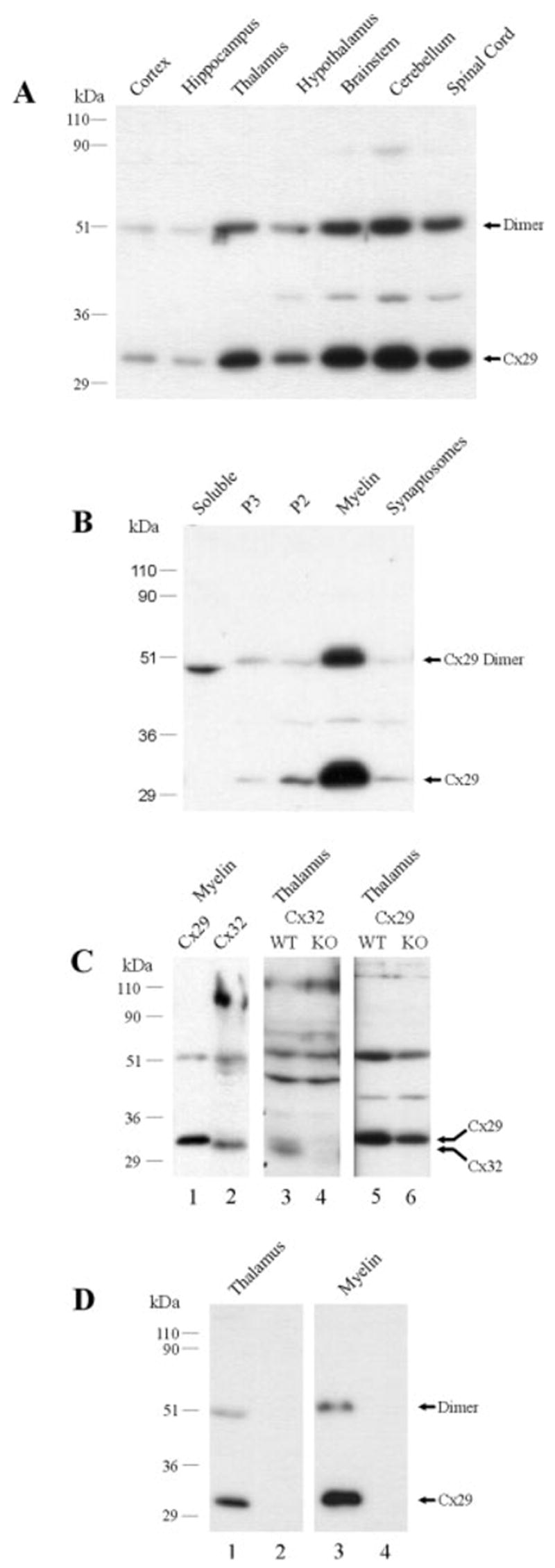
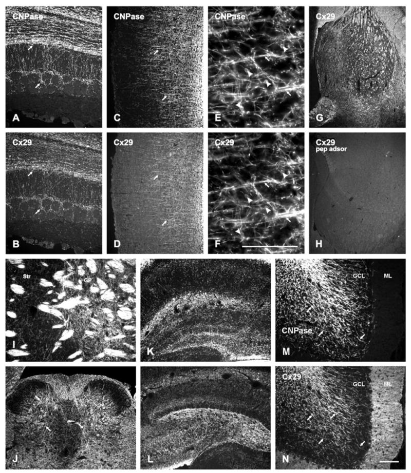
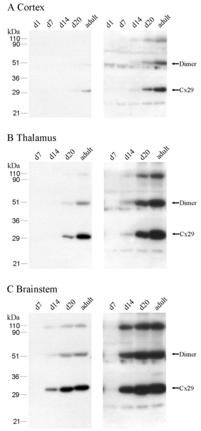
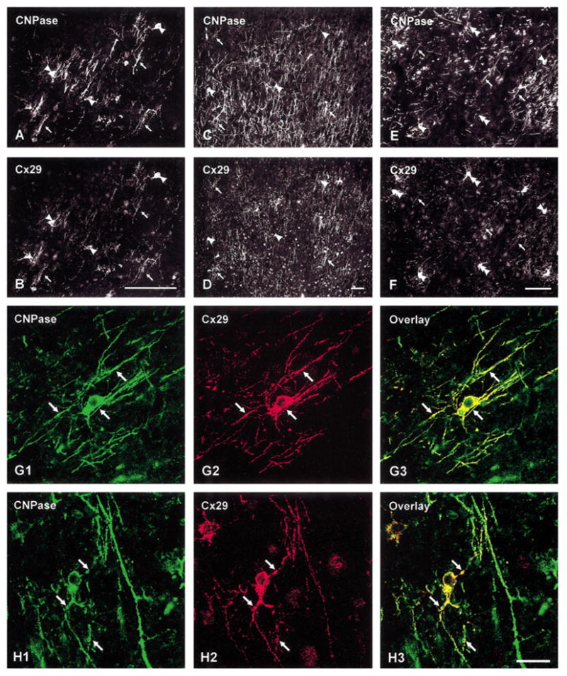
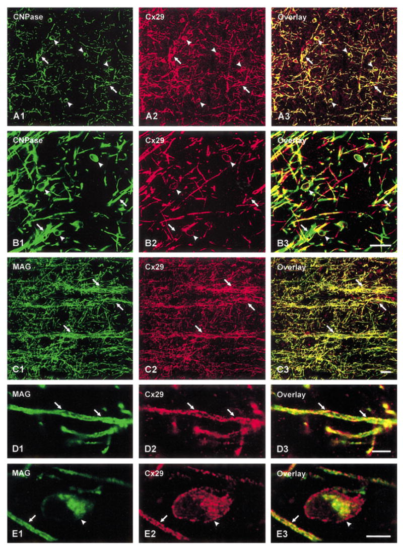
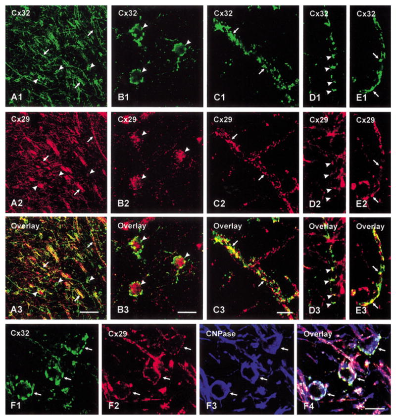
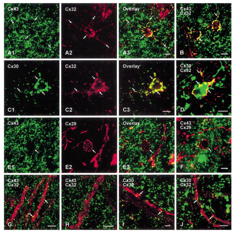
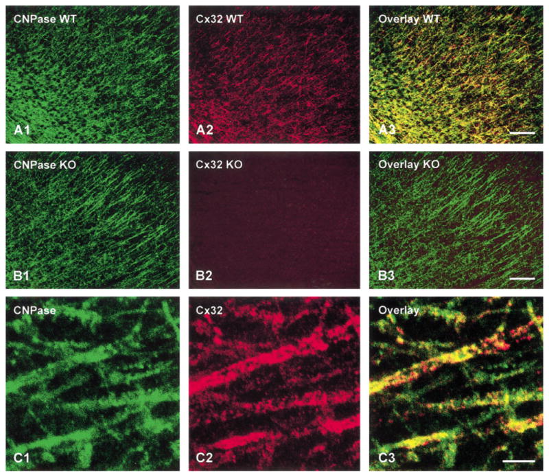
The cellular localization, relation to other glial connexins (Cx30, Cx32, and Cx43), and developmental expression of Cx29 were investigated in the mouse central nervous system (CNS) with an anti-Cx29 antibody. Cx29 was enriched in subcellular fractions of myelin, and immunofluorescence for Cx29 was localized to oligodendrocytes and myelinated fibers throughout the brain and spinal cord. Oligodendrocyte somata displayed minute Cx29-immunopositive puncta around their periphery and intracellularly. In developing brain, Cx29 levels increased during the first few postnatal weeks and were highest in the adult brain. Immunofluorescence labeling for Cx29 in oligodendrocyte somata was intense at young ages and was dramatically shifted in localization primarily to myelinated fibers in mature CNS. Labeling for Cx32 also was localized to oligodendrocyte somata and myelin and absent in Cx32 knockout mice. Cx29 and Cx32 were minimally colocalized on oligodendrocytes somata and partly colocalized along myelinated fibers. At gap junctions on oligodendrocyte somata, Cx43/Cx32 and Cx30/Cx32 were strongly associated, but there was minimal association of Cx29 and Cx43. Cx32 was very sparsely associated with astrocytic connexins along myelinated fibers. With Cx26, Cx30, and Cx43 expressed in astrocytes and Cx29, Cx32, and Cx47 expressed in oligodendrocytes, the number of connexins localized to gap junctions of glial cells is increased to six. The results suggested that Cx29 in mature CNS contributes minimally to gap junctional intercellular communication in oligodendrocyte cell bodies but rather is targeted to myelin, where it, with Cx32, may contribute to connexin-mediated communication between adjacent layers of uncompacted myelin.


Figures

Fig. 1
Fig. 1
Western blots of Cx29 and Cx32 in various neural tissues. In all blots, numbers at left correspond to molecular weight markers. A: Immunoblot showing regional levels of Cx29 in adult mouse brain. Monomeric and presumptive dimeric Cx29 are detected at 31 to 33 kDa and at 51 kDa, respectively. B: Immunoblot showing Cx29 in soluble, microsomal membrane (P3), mitochondrial/synaptosomal (P2), myelin, and synaptosomal subcellular fractions of adult brain. Cx29 is concentrated in the myelin fraction, and a protein of unknown identity at 48 kDa is detected in the soluble fraction. C: Immunoblots comparing Cx29 and Cx32 migration profiles. Lanes loaded with protein from myelin fraction and from thalamus homogenate of WT and Cx32 KO mice were probed with the antibodies indicated. Cx29 and Cx32 appear as distinct bands, and Cx32 at 30 to 31 kDa in the WT thalamus is absent in the Cx32 KO thalamus. Anti-Cx29 antibody detects Cx29 as corresponding bands in WT and Cx32 KO thalami. D: Immunoblots of the tissues indicated show elimination of Cx29 detection (lanes 2 and 4) after preadsorption of anti-Cx29 antibody with cognate peptide. Cx, connexin; KO, knockout; WT, wild type.
Fig. 2
Fig. 2
Immunofluorescence micrographs showing Cx29 localization in the CNS. A–D: Fields in olfactory bulb (A,B) and cerebral cortex (C,D) show overlapping distributions of fibers double labeled for CNPase (A,C, arrows) and Cx29 (B,D, arrows). E,F: Magnification of cerebral cortex showing colocalization of CNPase (E) and Cx29 (F) along myelinated fibers (arrows) and oligodendrocyte somata (arrowheads). G,H: Area in the thalamus (dorsal to the left) showing labeling for Cx29 (G) and elimination of labeling after preadsorption of anti-Cx29 with cognate peptide (H). I: Field at the border between the striatum (Str) and globus pallidus (GP) showing Cx29 in fiber bundles and in fibers scattered in neuropil of the GP. J: Lumbar spinal cord showing Cx29 in white matter and in myelinated fibers coursing through gray matter (arrows). K,L: Dentate gyrus of the hippocampus comparing labeling for Cx29 in sections from mouse (K) and rat (L) brains. M,N: Field in a cerebellar folia showing colocalization of CNPase (M) and Cx29 (N) along myelinated fibers (arrows) in white matter and in the granule cell layer (GCL). Dense immunoreactivity with anti-Cx29 is also seen in the molecular layer (ML), which is devoid of CNPase-positive fibers. CNPase, 2,′3′-cyclic nucleotide 3′-phosphodiesterase; Cx, connexin. Scale bars = 100 μm for E, F in and for A–D and G–N in N.
Fig. 3
Fig. 3
Western blots showing Cx29 levels in the cerebral cortex (A), thalamus (B), and brainstem (C) during development. Lanes were loaded with equal amounts of protein from homogenates of structures at the postnatal ages in days (d) indicated. For comparison of levels, blots are shown after exposure to film for short (left blot for each structure) and long (right blot for each structure) durations. Cx29 levels are highest in the adult brain, with increasing expression in each area during development. Cx, connexin.
Fig. 4
Fig. 4
Immunofluorescence micrographs illustrating developmental profiles of Cx29. A,B: Double labeling in a field of corpus callosum at postnatal day 8 showing CNPase-positive fibers (A, arrows) and oligodendrocyte somata (A, arrowheads) labeled for Cx29 (B). C–F: Double labeling for CNPase (C,E) and Cx29 (D,F) in a field of cerebral cortex (C,D) at postnatal day 14 and in the hypothalamus (E,F) at postnatal day 20. CNPase-positive fibers are sparsely labeled for Cx29 (arrows), whereas oligodendrocyte cell bodies (arrowheads) are intensely labeled for Cx29, but occasionally exhibit weak labeling for CNPase (E, double arrowheads). G,H: Confocal double immuno-fluorescence of fields in the cerebral cortex at postnatal day 14 showing colocalization of labeling for CNPase (G1,H1) and Cx29 (G2,H2) around an oligodendrocyte (G, arrows) and several Cx29-positive processes emanating from a Cx29-positive oligodendrocyte somata (H, arrows). G3, H3: Overlays. CNPase, 2,′3′-cyclic nucleotide 3′-phos-phodiesterase; Cx, connexin. Scale bars = 100 μm in A–F, 20 μm in G,H.
Fig. 5
Fig. 5
Confocal immunofluorescence showing association of Cx29 with the oligodendrocyte markers CNPase and MAG. A,B: Fields in the ventral anterior thalamic nucleus (A) and the cerebral cortex (B) double labeled for CNPase (A1,B1) and Cx29 (A2,B2). CNPase-positive fibers (arrows) and oligodendrocyte cell bodies (arrowheads) are immunopositive for Cx29 (yellow in image overlays, A3,B3). C–E: The same fields (C1,C2; D1,D2; and E1,E2) in the cerebral cortex double-labeled for MAG and Cx29, as indicated. Low and high magnifications show substantial colocalization of MAG and Cx29 along myelinated fibers (arrows) and partial colocalization in oligodendrocyte somata (E, arrowhead) as shown by yellow in image overlays (C3,D3,E3). Cx, connexin; MAG, myelin-associated glycoprotein. Scale bars = 20 μm in A–C, 2.5 μm in D,E.
Fig. 6
Fig. 6
Confocal immunofluorescence colocalization of Cx29 with Cx32 in the adult brain. Each column of images in A to E illustrates a field double labeled for Cx32 (green) and Cx29 (red), with colocalization appearing as yellow in image overlays. A: Low magnification in a field of thalamus showing Cx32 colocalization with Cx29 along myelinated fibers (arrows) and oligodendrocyte cell bodies (arrowheads). B: Oligodendrocyte somata in the thalamus displaying punctate labeling for Cx32 (B1, arrowheads), finer punctate labeling for Cx29 intracellularly and at the cell periphery (B2, arrowheads), and partial colocalization of the two connexins (B3). C–E: Labeling for Cx32 and Cx29 along myelinated fibers in the cerebral cortex (C,D) and thalamus (E) appears as puncta (C,E, arrows) or intermittent strands (D, arrowheads), which exhibit partial colocalization (C3,D3,E3). F: Confocal triple immunofluorescence showing that cells immunolabeled for Cx32 (F1, arrows) and Cx29 (F2, arrows) are CNPase positive (F3, arrows), as seen in overlay of images (F4). CNPase, 2,′3′-cyclic nucleotide 3′-phosphodiesterase; Cx, connexin. Scale bars = 20 μm in A, 10 μm in B, 5 μm in C3 for C–E, 5 μm in F.
Fig. 7
Fig. 7
A–C: Confocal double immunofluorescence of Cx43 and Cx30 with Cx32 in the adult brain. In fields of the hypothalamus (A) and thalamus (B; shown only by overlay), some Cx43-positive puncta are co-associated with Cx32 on oligodendrocytes and their initial processes (A,B, arrows). In a field of cerebral cortex (C), intense punctate labeling for Cx30 (C1, arrows) is co-associated with Cx32 (C2, arrows) on oligodendrocyte somata (C3, overlay). D: Confocal double labeling showing Cx30 association with CNPase-positive oligodendrocyte in the cerebral cortex (arrows) as seen by yellow in the overlay. E,F: Confocal double labeling of Cx43 and Cx29 in adult brain. In fields of globus pallidus (E) and hypothalamus (F; shown only by overlay), minimal co-association of Cx43-positive puncta is seen with Cx29 on oligodendrocyte somata (arrows). G–J: Confocal double immunofluorescence labeling of astrocytic Cx43 (green) and Cx30 (green) in relation to labeling of Cx32 (red) along myelinated fibers. Overlay images of fields in cerebral cortex (G) and thalamus (H) show limited co-association of Cx43-positive puncta with Cx32-positive fibers (arrows). Overlay images of fields in thalamus (I) and habenula (J) show sparse co-association of Cx30-positive puncta with Cx32 along fibers (arrows). CNPase, 2,′3′-cyclic nucleotide 3′-phosphodiesterase; Cx, connexin. Scale bars = 5 μm.
Fig. 8
Fig. 8
Immunofluorescence micrographs of Cx32 associated with myelinated fibers in the mouse cerebral cortex. A: Low magnification of a cortical field from a WT mouse double labeled for CNPase (A1) and Cx32 (A2), with colocalization shown in the overlay (yellow in A3). B: A cortical field from a Cx32 KO mouse double labeled for CNPase (B1) and Cx32 (B2) showing absence of Cx32 with consequent absence of yellow in the overlay (B3). C: Confocal double immunofluorescence of a field in a WT mouse showing CNPase-positive fibers (C1) labeled for Cx32 (C2) as seen by the overlay (C3). CNPase, 2,′3′-cyclic nucleotide 3′-phosphodiesterase; Cx, connexin; KO, knowckout; WT, wild type. Scale bars = 200 μm in A,B, 5 μm in C.

Similar articles

Cited by

References

    1. Altevogt BM, Kleopa KA, Postma FR, Scherer SS, Paul DL. Connexin29 is uniquely distributed within myelinating glial cells of the central and peripheral nervous systems. J Neurosci. 2002;22:6458–6470. - PMC - PubMed
    1. Arroyo EJ, Scherer SS. On the molecular architecture of myelinated fibres. Histochem Cell Biol. 2000;113:1–18. - PubMed
    1. Balice-Gordon RJ, Bone LJ, Scherer SS. Functional gap junctions in the Schwann cell myelin sheath. J Cell Biol. 1998;142:1095–1104. - PMC - PubMed
    1. Bergoffen J, Scherer SS, Wang S, Oronzi-Scott M, Bone L, Paul DL, Chen K, Lensch MW, Chance P, Fischbeck K. Connexin mutations in X-linked Charcot-Marie-Tooth disease. Science. 1993;262:2039 –2042. - PubMed
    1. Bone LJ, Deschênes SM, Balice-Gordon RJ, Fischbeck KH, Scherer SS. Connexin 32 and X-linked Charcot-Marie-Tooth disease. Neurobiol Dis. 1997;4:221–230. - PubMed

Publication types

MeSH terms