. 2003 Aug;69(8):4915-26.
doi: 10.1128/AEM.69.8.4915-4926.2003.

Colonization of Arabidopsis thaliana with Salmonella enterica and enterohemorrhagic Escherichia coli O157:H7 and competition by Enterobacter asburiae

Affiliations

Colonization of Arabidopsis thaliana with Salmonella enterica and enterohemorrhagic Escherichia coli O157:H7 and competition by Enterobacter asburiae

Michael B Cooley et al. Appl Environ Microbiol. 2003 Aug.

Abstract

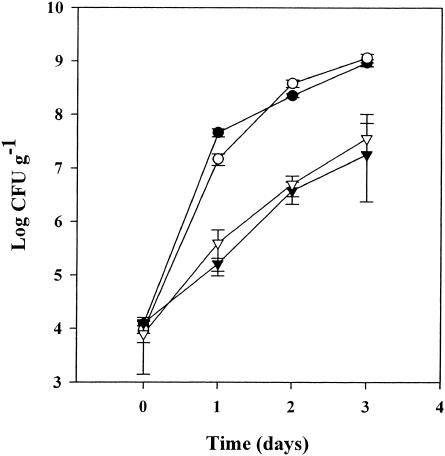
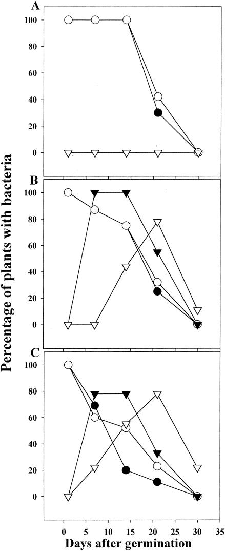
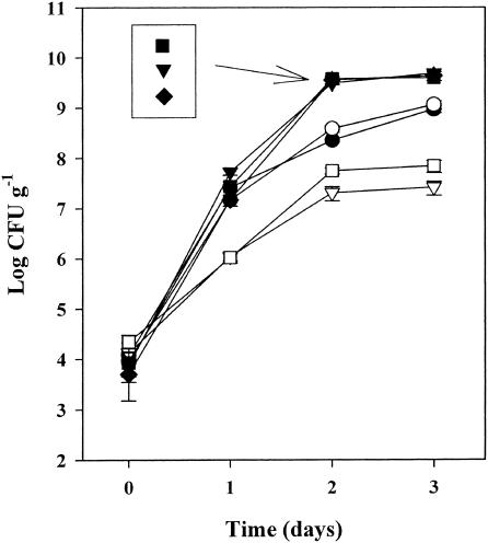
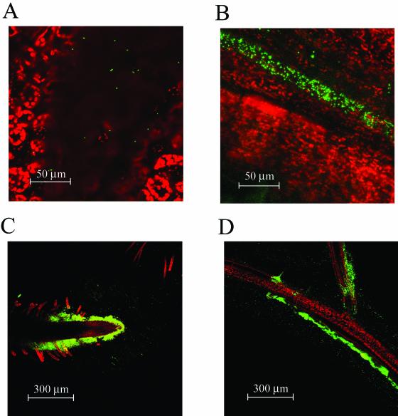
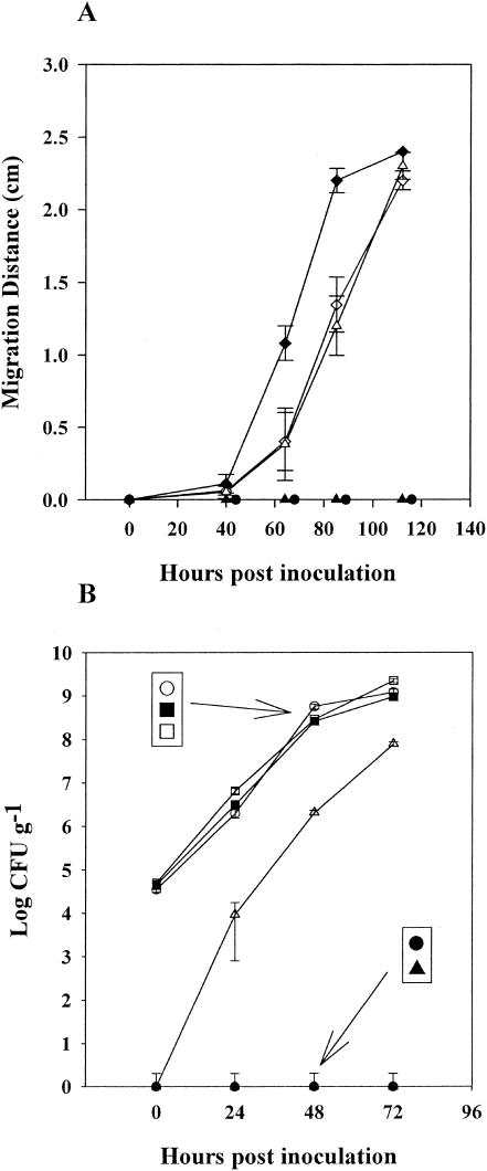
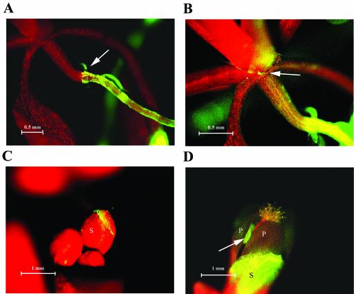
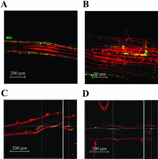
Enteric pathogens, such as Salmonella enterica and Escherichia coli O157:H7, have been shown to contaminate fresh produce. Under appropriate conditions, these bacteria will grow on and invade the plant tissue. We have developed Arabidopsis thaliana (thale cress) as a model system with the intention of studying plant responses to human pathogens. Under sterile conditions and at 100% humidity, S. enterica serovar Newport and E. coli O157:H7 grew to 10(9) CFU g(-1) on A. thaliana roots and to 2 x 10(7) CFU g(-1) on shoots. Furthermore, root inoculation led to contamination of the entire plant, indicating that the pathogens are capable of moving on or within the plant in the absence of competition. Inoculation with green fluorescent protein-labeled S. enterica and E. coli O157:H7 showed invasion of the roots at lateral root junctions. Movement was eliminated and invasion decreased when nonmotile mutants of S. enterica were used. Survival of S. enterica serovar Newport and E. coli O157:H7 on soil-grown plants declined as the plants matured, but both pathogens were detectable for at least 21 days. Survival of the pathogen was reduced in unautoclaved soil and amended soil, suggesting competition from indigenous epiphytes from the soil. Enterobacter asburiae was isolated from soil-grown A. thaliana and shown to be effective at suppressing epiphytic growth of both pathogens under gnotobiotic conditions. Seed and chaff harvested from contaminated plants were occasionally contaminated. The rate of recovery of S. enterica and E. coli O157:H7 from seed varied from undetectable to 19% of the seed pools tested, depending on the method of inoculation. Seed contamination by these pathogens was undetectable in the presence of the competitor, Enterobacter asburiae. Sampling of 74 pools of chaff indicated a strong correlation between contamination of the chaff and seed (P = 0.025). This suggested that contamination of the seed occurred directly from contaminated chaff or by invasion of the flower or silique. However, contaminated seeds were not sanitized by extensive washing and chlorine treatment, indicating that some of the bacteria reside in a protected niche on the seed surface or under the seed coat.


Figures

FIG. 1.
FIG. 1.
Growth of S. enterica serovar Newport (○, ▿) and E. coli O157:H7 (•, ▾) on A. thaliana shoots (▾, ▿) and roots (•, ○). Inoculation and growth of the bacteria were performed with the roots exposed on the surface of agar plates. Bacterial concentrations are averages of six samples. Error bars show standard errors.
FIG. 2.
FIG. 2.
Incidence of recovery of S. enterica serovar Newport (○) and E. coli O157:H7 (•) from plants grown in autoclaved (A), unautoclaved (B), and amended (C) soils and coincident isolation of Enterobacter asburiae from S. enterica serovar Newport-infected (▿) and E. coli O157:H7-infected (▾) plants. Each data point represents samples from 36 plants.
FIG. 3.
FIG. 3.
Growth of Enterobacter asburiae (▾), S. enterica serovar Newport (•), and E. coli O157:H7 (○) separately on roots of sterilely grown plants or coinoculated as S. enterica serovar Newport (□) or E. coli O157:H7 (▿) with Enterobacter asburiae (▪ ♦, respectively). Bacterial concentrations are averages of six samples. Error bars show standard errors.
FIG. 4.
FIG. 4.
Colonization of A. thaliana leaves by GFP-labeled E. coli O157:H7 localized in small depressions (A) and over veins (B). Chloroplast autofluorescence is colored red. Initial colonization of roots occurs at root tips (C) and the branch points of lateral roots (D). Red indicates either vascular autofluorescence or TO-PRO-3 staining of epidermis and root hairs (see Materials and Methods).
FIG. 5.
FIG. 5.
Growth of bacteria and migration along the root following inoculation 2.4 cm below the crown. (A) Migration of GFP-labeled E. coli O157:H7 (♦), S. enterica serovar Typhimurium SJW1103 (▵), S. enterica serovar Newport (⋄), S. enterica serovar Typhimurium SJW1368 (•), and S. enterica serovar Typhimurium SJW1809 (▴) above the region of inoculation was observed by stereo epifluorescence microscope. Migration distance is the extent of bacterial movement during the elapsed time and is the average of 10 measurements on different plants. (B) Growth of GFP-labeled S. enterica serovar Typhimurium SJW1103, S. enterica serovar Typhimurium SJW1368 and S. enterica serovar Typhimurium SJW1809 in the inoculated region of the root (○, ▪, and □, respectively) and above the inoculated region (▵, •, and ▴, respectively). Bacterial concentrations are averages of six samples. Error bars show standard errors.
FIG. 6.
FIG. 6.
Migration of GFP-labeled E. coli O157:H7 from the crown to the flowers. (A) Bacteria have moved up the root from the lower right to the crown (indicated by the arrow). (B) Longer exposure of the meristematic region shown in panel A, showing colonization at the base of the petioles (indicated by the arrow). (C) Colonization of three floral buds. (D) Colonization of the sepal of an opening flower. Note: green autofluorescence of the anther (indicated by the arrow) was also observed in flowers of uninoculated plants. The red in panels A to D is chloroplast autofluorescence. S, sepal; P, petal.
FIG. 7.
FIG. 7.
Invasion of GFP-labeled bacteria into the primary root at lateral root junctions. (A) Invasion of S. enterica serovar Newport at a newly developed lateral root that has just broken the epidermis of the primary root. Fluorescent bacteria are seen just below and adjacent to the epidermal cells. (B) S. enterica serovar Newport within the primary root appears to occupy the intercellular regions (apoplast). The bacteria delineate cells within the primary root. (C) E. coli O157:H7 within the primary root at a mature lateral root junction. The right panel is a computer-generated vertical slice at the line in the left panel, showing that the bacteria are within the root, not on the surface. The lateral root is out of the focal plane. (D) E. coli O157:H7 invasion of a mature lateral root, similar to panel C but showing greater spread of the bacteria along the primary root. The red in panels A to D is either vascular autofluorescence or TO-PRO-3 staining of epidermis and root hairs (see Materials and Methods).

Similar articles

Cited by

References

    1. Anonymous. 1997. Outbreaks of Escherichia coli O157:H7 infection and cryptosporidiosis associated with drinking unpasteurized apple cider—Connecticut and New York, October 1996. Morbid. Mortal. Wkly. Rep. 46:4-8. - PubMed
    1. Anonymous. 1996. Standardized molecular subtyping of foodborne bacterial pathogens by pulsed-field gel electrophoresis: a manual. National Center for Infectious Diseases, Centers for Disease Control and Prevention, Atlanta, Ga.
    1. Arora, D. K., A. B. Filonow, and J. L. Lockwood. 1983. Bacterial chemotaxis to fungal propagules in vitro and in soil. Can. J. Microbiol. 29:1104-1109.
    1. Baloda, S. B., L. Christensen, and S. Trajcevska. 2001. Persistence of a Salmonella enterica serovar typhimurium DT12 clone in a piggery and in agricultural soil amended with Salmonella-contaminated slurry. Appl. Environ. Microbiol. 67:2859-2862. - PMC - PubMed
    1. Bayot, R. G., and S. M. Ries. 1986. Role of motility in apple blossom infection by Erwinia amylovora and studies of fire blight control with attractant and repellent compounds. Phytopathology 76:441-445.

Publication types

MeSH terms

LinkOut - more resources