Comparative Study
. 2003 Aug 15;171(4):2099-108.
doi: 10.4049/jimmunol.171.4.2099.

Intracellular domain of brain endothelial intercellular adhesion molecule-1 is essential for T lymphocyte-mediated signaling and migration

Affiliations
Comparative Study

Intracellular domain of brain endothelial intercellular adhesion molecule-1 is essential for T lymphocyte-mediated signaling and migration

John Greenwood et al. J Immunol. .

Abstract

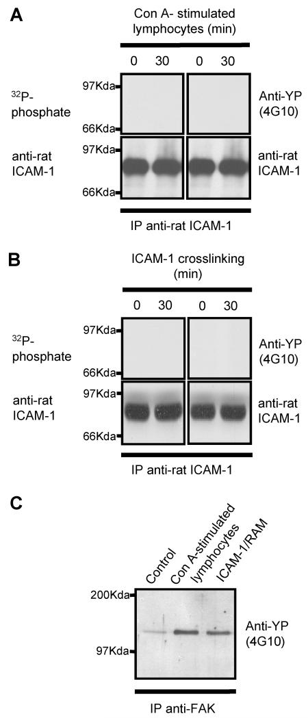
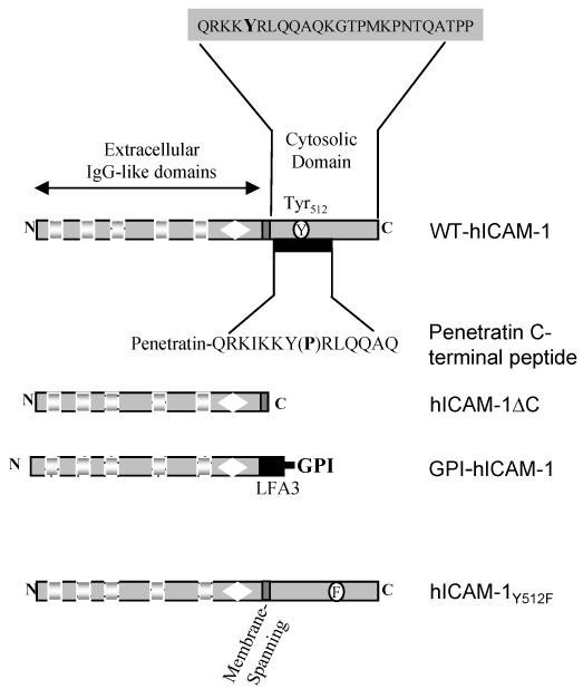
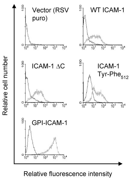
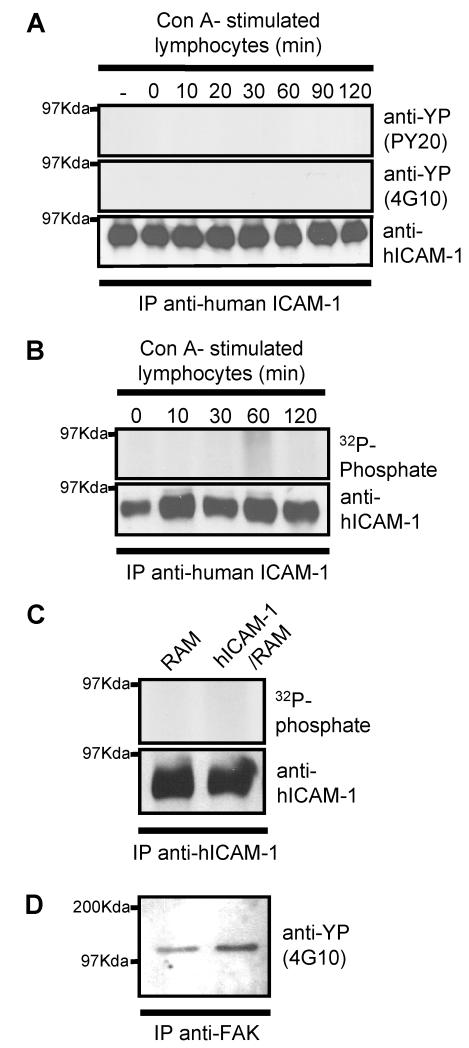
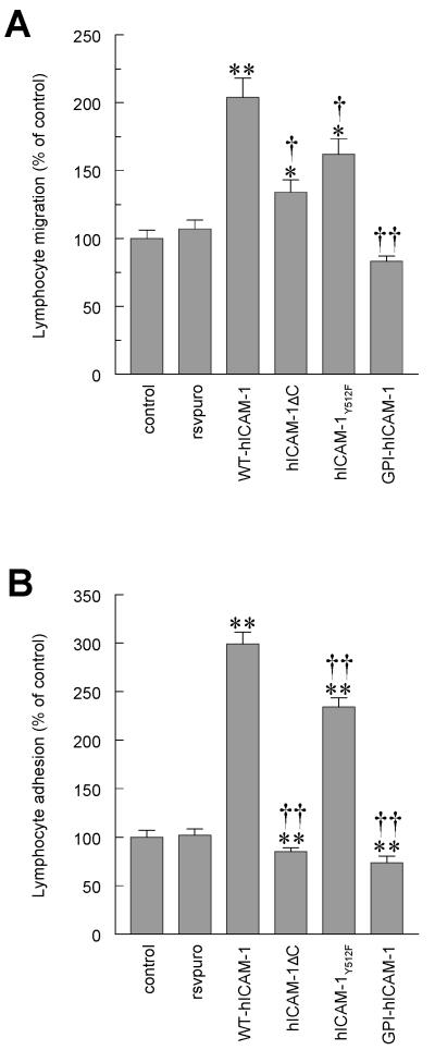
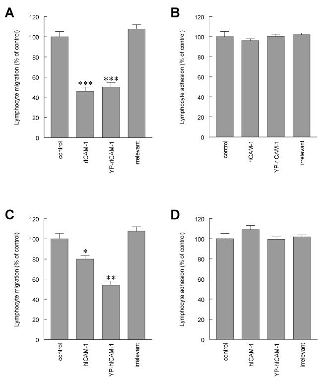
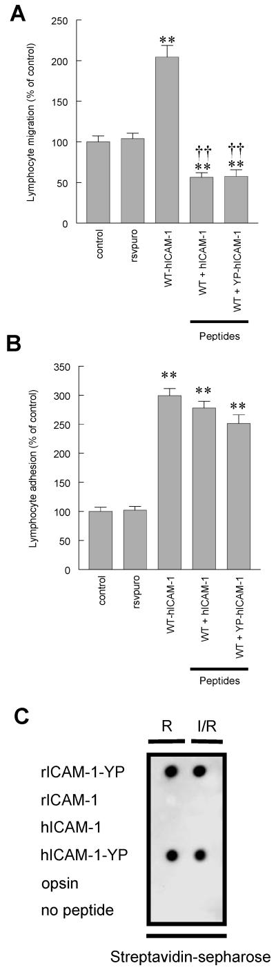
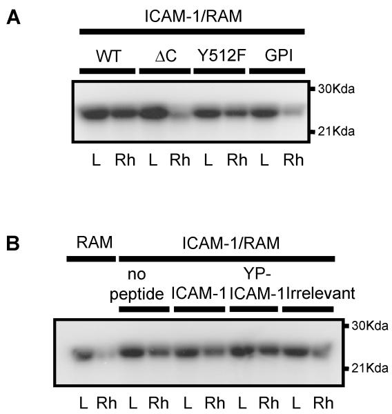
To examine the role of the ICAM-1 C-terminal domain in transendothelial T lymphocyte migration and ICAM-1-mediated signal transduction, mutant human (h)ICAM-1 molecules were expressed in rat brain microvascular endothelial cells. The expression of wild-type hICAM-1 resulted in a significant increase over basal levels in both adhesion and transendothelial migration of T lymphocytes. Endothelial cells (EC) expressing ICAM-1 in which the tyrosine residue at codon 512 was substituted with phenylalanine (hICAM-1(Y512F)) also exhibited increased lymphocyte migration, albeit less than that with wild-type hICAM-1. Conversely, the expression of truncated hICAM-1 proteins, in which either the intracellular domain was deleted (hICAM-1DeltaC) or both the intracellular and transmembrane domains were deleted through construction of a GPI anchor (GPI-hICAM-1), did not result in an increase in lymphocyte adhesion, and their ability to increase transendothelial migration was attenuated. Truncated hICAM-1 proteins were also unable to induce ICAM-1-mediated Rho GTPase activation. EC treated with cell-permeant penetratin-ICAM-1 peptides comprising human or rat ICAM-1 intracellular domain sequences inhibited transendothelial lymphocyte migration, but not adhesion. Peptides containing a phosphotyrosine residue were equipotent in inhibiting lymphocyte migration. These data demonstrate that the intracellular domain of ICAM-1 is essential for transendothelial migration of lymphocytes, and that peptidomimetics of the ICAM-1 intracellular domain can also inhibit this process. Such competitive inhibition of transendothelial lymphocyte migration in the absence of an affect on adhesion further implicates ICAM-1-mediated signaling events in the facilitation of T lymphocyte migration across brain EC. Thus, agents that mimic the ICAM-1 intracellular domain may be attractive targets for novel anti-inflammatory therapeutics.


Figures

Figure 1
Figure 1. Endogenous endothelial rat ICAM-1 is not phosphorylated following co-culture with T-lymphocytes or cross-linking of endothelial ICAM-1
Unlabelled or [32P]-labelled rat brain EC were either (A) co-cultured for various times with concanavalin-A stimulated peripheral lymph node lymphocytes or (B) crosslinked with anti-rat ICAM-1 followed by rabbit anti-mouse IgG (RAM). EC were washed and ICAM-1 or FAK immunoprecipitated from EC lysates using anti-rat ICAM-1 (1A29) or anti-FAK. ICAM-1 immunoprecipitates from [32P]-labelled cells were resolved on 10% SDS-PAGE and exposed to autoradiographic film or immunoblotted with anti-phosphotyrosine mAb (4G10). Membranes were subsequently stripped and re-probed for rat ICAM-1 using 1A29 mAb. FAK immunoprecipitates (C) were immunoblotted with anti-phosphotyrosine mAb (4G10).
Figure 2
Figure 2. Schematic illustration of wild type and mutant human ICAM-1 molecules and targeted peptide sequences within the cytoplasmic domain of ICAM-1
Wild type human ICAM-1 (WT-hICAM-1); human ICAM-1 with 28 residue C-terminal truncation (hICAM-1ΔC); human ICAM-1 in which the cytoplasmic and transmembrane domains have been removed and which has subsequently been fused to the GPI-linker region of human LFA3 (GPI-hICAM-1); human ICAM-1 carrying a tyrosine to phenylalanine substitution at codon 512 (hICAM-1Y512F).
Figure 3
Figure 3. Flow cytometric analysis of rat brain EC expressing wild type and mutant human ICAM-1 molecules
Cell lines transfected with human ICAM-1 constructs were stained with mouse anti-human ICAM-1 (BBA4; black histogram) or isotype matched control IgG (grey histogram) followed by anti-mouse-FITC (1:50). 10,000 events were acquired and histograms show analysis of human ICAM-1 expression of live cell population.
Figure 4
Figure 4. Human ICAM-1 is not phosphorylated following co-culture of rat brain EC with T-lymphocytes or cross-linking with human ICAM-1
Unlabelled or [32P]-labelled brat rain EC expressing human wild type ICAM-1 were co-cultured for various times with concanavalin-A stimulated peripheral lymph node lymphocytes. Cells were washed and human ICAM-1 immunoprecipitated from EC lysates using anti-human ICAM-1 mAb (BBA4). Immunoprecipitates were resolved on SDS-PAGE, transferred to nitrocellulose membranes, and immunoblotted with (A) anti-phosphotyrosine clone PY20 (upper panel) and clone 4G10 (middle panel) or immunoblotted with anti-human ICAM-1 (lower panel.) (B) Exposed to autoradiographic film (upper panel) or immunoblotted with anti-human ICAM-1 (lower panel.). (C) [32P]-labelled rat brain EC, expressing wild type human ICAM-1, were cross-linked with anti-human ICAM-1 mAb (BBA4) followed by rabbit anti-mouse IgG (RAM). Human ICAM-1 or rat FAK was immunoprecipitated with BBA4 or anti-FAK. ICAM-1 immunoprecipitates were resolved on SDS-PAGE and exposed to autoradiographic film (upper panel) or immunoblotted with anti-human ICAM-1 (lower panel.). (D) FAK immunoprecipitates from human ICAM-1 cross-linked cells immunoblotted with anti-phosphotyrosine mAb (4G10)(lower panel).
Figure 5
Figure 5. Lymphocyte adhesion to and migration through rat brain EC monolayers expressing human ICAM-1
(A) Migration of antigen specific (MBP) T cells through or (B) adhesion of [3H]-labelled activated rat peripheral lymph node lymphocytes to control EC monolayers, EC transfected with RSVpuro empty vector (rsvpuro), EC expressing human wild type ICAM-1 (WT-hICAM-1), EC expressing ICAM-1 truncated at codon 504 (hICAM-1ΔC), EC expressing human ICAM-1 with tyrosine to phenylalanine substitution at codon 512 (hICAM-1Y512F) or EC expressing human ICAM-1 truncated at amino acid 480 and fused to the glycophosphatidyl inositol linker of LFA3 (GPI-hICAM-1). Data are expressed as Mean ± SEM percent of control of a minimum of three independent experiments (n= >6 per experiment). Significant differences were determined by Student’s t-test. Compared to control (RSVpuro vector) *p<0.01, **p<0.001 or compared to Wild Type human ICAM-1 † <0.02, † † p<0.001, Student t-test.
Figure 6
Figure 6. Lymphocyte adhesion to and migration through rat brain EC monolayers in the presence of human and rat penetratin-ICAM-1 peptides
(A and C) Migration of antigen specific (MBP) T cells through or (B and D) adhesion of [3H]-labelled activated rat peripheral lymph node lymphocytes to control rat brain EC monolayers or EC pretreated with rat (A and B) or human (C and D) penetratin peptides. Penetratin peptide containing C-terminal 16 amino acids of rat ICAM-1 (rICAM-1), penetratin peptide containing C-terminal 16 amino acids of rat ICAM-1 in which the tyrosine residue is phosphorylated (YP-rICAM-1), penetratin peptide containing C-terminal 16 amino acids of human ICAM-1 (hICAM-1), penetratin peptide containing C-terminal 16 amino acids of human ICAM-1 in which the tyrosine residue is phosphorylated (YP-hICAM-1) and penetratin peptide containing 16 amino acids or rat opsin (irrelevant). Observations are a minimum of three independent experiments (n=6/experiment). Data are expressed as Mean ± SEM percent of control of a minimum of three independent experiments (n= >6 per experiment). Significant differences compared to control were determined by Student’s t-test *p<0.01, **p<0.001, ***p<0.0001
Figure 7
Figure 7. Lymphocyte adhesion to and migration through rat brain EC monolayers expressing human ICAM-1: Effect of penetratin-human ICAM-1 peptides
(A) Migration of antigen specific (MBP) T cells through or (B) adhesion of [3H]-labelled activated rat peripheral lymph node lymphocytes to control EC monolayers, EC transfected with RSVpuro empty vector (rsvpuro) or EC expressing human wild type ICAM-1 (WT-hICAM-1). EC expressing WT-hICAM-1 were also pretreated with either penetratin peptide containing C-terminal 16 amino acids of human ICAM-1 (hICAM-1) or penetratin peptide containing C-terminal 16 amino acids of ICAM-1 in which the tyrosine residue is phosphorylated (YP-hICAM-1). Observations are a minimum of three independent experiments (n=6/experiment). Data are expressed as Mean ± SEM percent of control of a minimum of three independent experiments (n= >6 per experiment). Significant differences were determined by Student’s t-test. **p<0.001 compared to wild type human ICAM-1. † † p<0.001 compared to RSVpuro control. (C) Anti-phosphotyrosine dot-blot analysis of biotinylated penetratin-hICAM-1 peptides extracted with streptavidin-spepharose from RAM (R) only and ICAM-1/RAM (I/R) treated GP8/3.9 cells expressing WT-hICAM-1.
Figure 8
Figure 8
(A) Rat brain EC expressing human ICAM-1 constructs or (B) rat brain EC expressing WT human ICAM-1 in the presence of C-terminal ICAM-1 penetratin peptides were cross linked with anti-human ICAM-1 followed by rabbit anti-mouse IgG (RAM) or RAM only for 15 min, lysed and activated Rho proteins precipitated with GST-rhotekin. Total Rho proteins and GST-rhotekin precipitated Rho proteins were subsequently [32P]ADP-ribosylated, resolved on 15%SDS-PAGE and exposed to autoradiographic film. L = lysate, Rh = precipitated GST-rhotekin. (C) Rat brain EC treated with anti-rat penetratin ICAM-1 peptides (100μg/ml, 2 h) and subsequently cross-linked with anti-rat ICAM-1 for 30 min followed by RAM for 15 min. Cells were fixed and F-actin and biotinylated peptides detected by direct immunofluoresence. (a) penetratin-hICAM-1, (b) F-actin and (c) merge of a and b. (d) penetratin-YP-hICAM-1, (e) F-actin and (f) merge of d and e. (g) penetratin-opsin, (h) F-actin and (i) merge of g and h. Scale bars = 25 μm. (D) Brain EC microinjected in the cytosol with biotinylated penetratin-peptides (10 mg/ml). (a) Cells stained for biotiylated-YP-hICAM-1 peptide. (b) identical field to (a) showing F-actin. (c) merge of (a) and (b). (d and e) merged images of cells microinjected with biotinylated hICAM-1 peptide and (f) biotinylated penetratin-opsin. Scale bars: c and d = 50 μm; e and f = 20 μm
Figure 8
Figure 8
(A) Rat brain EC expressing human ICAM-1 constructs or (B) rat brain EC expressing WT human ICAM-1 in the presence of C-terminal ICAM-1 penetratin peptides were cross linked with anti-human ICAM-1 followed by rabbit anti-mouse IgG (RAM) or RAM only for 15 min, lysed and activated Rho proteins precipitated with GST-rhotekin. Total Rho proteins and GST-rhotekin precipitated Rho proteins were subsequently [32P]ADP-ribosylated, resolved on 15%SDS-PAGE and exposed to autoradiographic film. L = lysate, Rh = precipitated GST-rhotekin. (C) Rat brain EC treated with anti-rat penetratin ICAM-1 peptides (100μg/ml, 2 h) and subsequently cross-linked with anti-rat ICAM-1 for 30 min followed by RAM for 15 min. Cells were fixed and F-actin and biotinylated peptides detected by direct immunofluoresence. (a) penetratin-hICAM-1, (b) F-actin and (c) merge of a and b. (d) penetratin-YP-hICAM-1, (e) F-actin and (f) merge of d and e. (g) penetratin-opsin, (h) F-actin and (i) merge of g and h. Scale bars = 25 μm. (D) Brain EC microinjected in the cytosol with biotinylated penetratin-peptides (10 mg/ml). (a) Cells stained for biotiylated-YP-hICAM-1 peptide. (b) identical field to (a) showing F-actin. (c) merge of (a) and (b). (d and e) merged images of cells microinjected with biotinylated hICAM-1 peptide and (f) biotinylated penetratin-opsin. Scale bars: c and d = 50 μm; e and f = 20 μm
Figure 9
Figure 9. Both WT-hICAM-1 and hICAM-1ΔC form rat:human heterodimers
(A) Rat brain EC cells expressing human ICAM-1 or hICAM-1ΔC ere treated with varying concentrations of the reversible cross-linker DTSSP. Cells were lysed and proteins resolved on 6% non-reducing SDS-PAGE prior to immunoblotting with anti-human ICAM mAb (BBA4). Arrows denote ICAM-1 and ICAM-1 dimers. L = lysate of cells with no cross-linker. (B) Cells expressing WT human ICAM-1 or hICAM-1ΔC were immunoprecipitated using anti-rat ICAM-1 mAb (1A29) and immunoprecipitates immunoblotted with anti-human ICAM-1 mAb (BBA4). (C) Cells expressing WT human ICAM-1 were immunoprecipitated with anti-human ICAM-1 mAb (BBA4) and immunoblotted with anti-rat ICAM-1 mAb (1A29). (D) brain EC lysate immunoblotted with anti-human ICAM-1 mAb (BBA4) demonstrating no cross-reactivity with rat ICAM-1.

Similar articles

Cited by

References

    1. Male D, Pryce G, Hughes C, Lantos P. Lymphocyte migration into brain modeled in vitro: control by lymphocyte activation, cytokines, and antigen. Cell. Immunol. 1990;127:1. - PubMed
    1. Greenwood J, Wang Y, Calder VL. Lymphocyte adhesion and transendothelial migration in the central nervous system: the role of LFA-1, ICAM-1, VLA-4 and VCAM-1. Immunology. 1995;86:408. - PMC - PubMed
    1. Pryce G, Male DK, Campbell I, Greenwood J. Factors controlling T-cell migration across rat cerebral endothelium in vitro. J. Neuroimmunol. 1997;75:84. - PubMed
    1. Etienne S, Adamson P, Greenwood J, Strosberg AD, Cazaubon S, Couraud P-O. Rho-dependent signaling pathways coupled to ICAM-1 in microvascular brain endothelial cells. J. Immunol. 1998;161:5755. - PubMed
    1. Adamson P, Etienne S, Couraud P-O, Calder V, Greenwood J. Lymphocyte migration through brain endothelial cell monolayers involves signalling through endothelial ICAM-1 via a Rho-dependent pathway. J. Immunol. 1999;162:2964. - PubMed

Publication types

MeSH terms

Substances