. 2003 Aug 19;100(17):9946-51.
doi: 10.1073/pnas.1334037100. Epub 2003 Aug 5.

Proteasome inhibitors disrupt the unfolded protein response in myeloma cells

Affiliations

Proteasome inhibitors disrupt the unfolded protein response in myeloma cells

Ann-Hwee Lee et al. Proc Natl Acad Sci U S A. .

Abstract

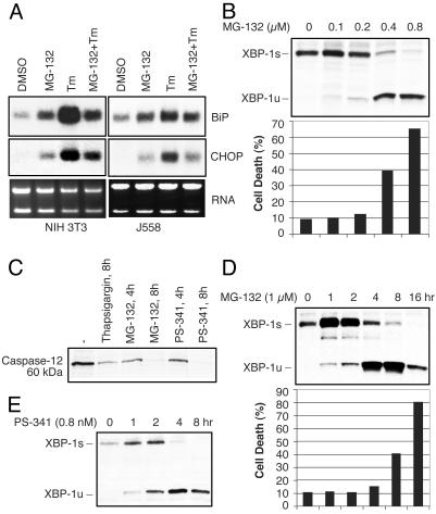
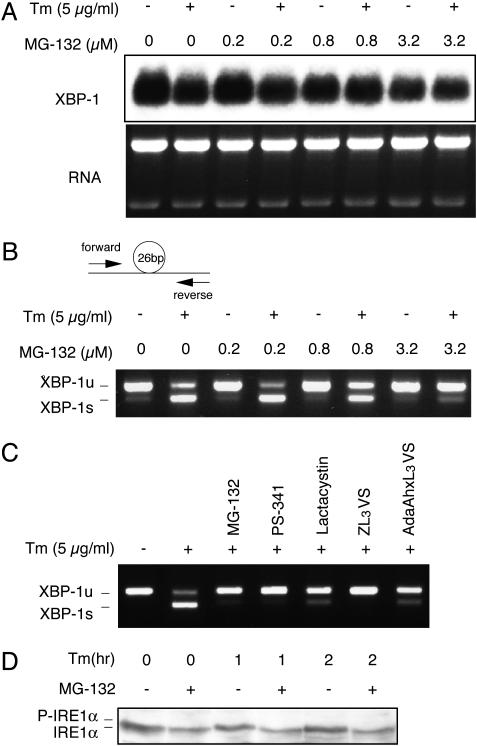
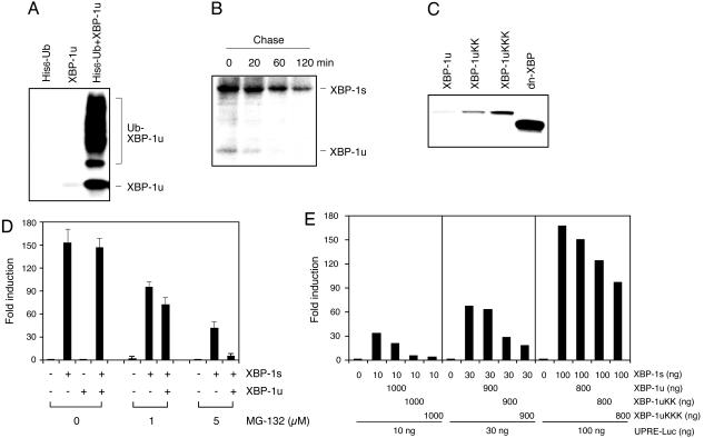
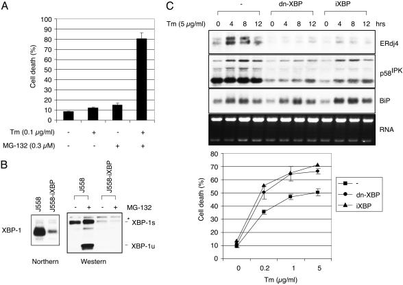
Novel agents that target the proteasome, a proteolytic complex responsible for the degradation of ubiquitinated proteins, have demonstrated remarkable therapeutic efficacy in multiple myeloma, a plasma cell malignancy. However, the mechanism by which these compounds act remains unknown. A signaling pathway called the unfolded protein response (UPR) allows cells to handle the proper folding of proteins. The transcription factor XBP-1, a regulator of the UPR, is also required for plasma cell differentiation, suggesting a link between the UPR and plasma cell differentiation. Here we show that proteasome inhibitors target XBP-1 and the UPR in myeloma cells. Proteasome inhibitors suppress the activity of the translumenal endoplasmic reticulum endoribonuclease/kinase, IRE1 alpha, to impair the generation of the active, spliced XBP-1 species and simultaneously stabilize the unspliced species that acts as a dominant negative. Myeloma cells rendered functionally deficient in XBP-1 undergo increased apoptosis in response to endoplasmic reticulum stress. Identification of compounds that target the activity of IRE1 alpha/XBP-1 may yield novel therapies for the treatment of multiple myeloma and other malignancies that rely on an intact UPR.


Figures

Fig. 1.
Fig. 1.
PIs induce ER stress and caspase-12 activation, but suppress the UPR. (A) BiP and CHOP mRNA induction in NIH 3T3 or J558 myeloma cells treated with MG-132 (20 μM), Tm (10 μg/ml), or both. Cells were pretreated with MG-132 for 1 h and then further treated with Tm for 4 h. Ethidium bromide staining of the gel is shown at the bottom. (B) Alteration in the ratio of XBP-1 protein species in J558 cells treated with increasing amounts of MG-132 for 16 h. Cells undergoing apoptosis were counted by annexin V staining. (C) Inhibition of caspase-12 processing by PIs. Processing of full-length caspase-12 was examined by Western blotting in J558 myeloma cells treated with thap-sigargin (1 μM) or PIs (20 μM) during the indicated time periods. (D) Time course of the XBP-1s to -1u shift. Cells were treated with MG-132 (1 μM) for the indicated times, and XBP-1u and -1s protein levels and cell death were determined. (E) Alteration in the ratio of XBP-1 protein species in the MM.1s human myeloma cell line. Cells were treated with PS-341 (8 nM) in a time course analysis, and XBP-1 protein species were quantified.
Fig. 2.
Fig. 2.
Effect of PIs on IRE1α-mediated XBP-1 mRNA splicing. (A) XBP-1 mRNA levels in ER-stressed J558 cells treated with Tm for4hinthe absence or presence of MG-132. Cells were pretreated with MG-132 for 1 h before adding Tm. (B) The ratio of XBP-1u to -1s mRNA as revealed by RT-PCR analysis with a probe set spanning the spliced-out region as demonstrated (22). (C) Effect of a panel of PIs on XBP-1 splicing. Cells were treated with Tm for 4 h in the absence or presence of MG-132 (10 μM), PS-341 (10 μM), lactacystin (10 μM), ZL3VS (50 μM), or AdaAhxL3VS (50 μM). (D) IRE1α phosphorylation in NIH 3T3 cells treated with Tm as indicated after 2 h of pretreatment with MG-132 (10 μM).
Fig. 3.
Fig. 3.
PIs stabilize XBP-1u protein to act as a dominant-negative inhibitor of XBP-1s activity. (A) Ubiquitination of XBP-1 in HeLa cells cotransfected with XBP-1u and His-tagged ubiquitin expression plasmids. (B) Degradation rates of XBP-1u and -1s proteins as determined by pulse-labeling J558 cells with [35S]Met/[35S]Cys for 1 h and chasing for the indicated times. (C) Generation and expression of lysine to arginine XBP-1u mutants. Two or three lysine residues in the C terminus of XBP-1u were replaced by arginine to generate XBP-1uKK (235, 252) and XBP-1uKKK (146, 235, 252) by site-directed mutagenesis. dn-XBP contains the N-terminal 188 aa of XBP-1u. Western blot analysis was performed with NIH 3T3 extracts transfected with the indicated plasmids. (D) Effect of XBP-1u on XBP-1s-dependent UPR element (UPRE) activation in PI-treated NIH 3T3 cells with 8-fold excess of XBP-1u plasmids. Transfected cells were treated with MG-132 for 16 h before harvesting for luciferase assays. Values represent fold induction of activity compared with the reporter alone after normalizing to Renilla. (E) Inhibition of XBP-1s-dependent activation of the UPRE reporter in NIH 3T3 cells by XBP-1u lysine to arginine mutants.
Fig. 4.
Fig. 4.
Cells with an impaired UPR are more sensitive to ER stress-induced apoptosis. (A) Synergistic effect of Tm and MG-132 on apoptosis. Annexin V-positive cells were counted after treating J558 cells for 18 h with suboptimal concentrations of Tm and MG-132 as indicated. (B) Generation of J558-iXBP cells by retroviral transduction of J558 cells with the U6 promoter-based XBP-1 RNAi vector. (C) XBP-1-dependent gene expression in J558 cells that express control GFP, dn-XBP, or iXBP-1 treated with Tm. Generation of dn-XBP-1 J558 cells by infection with a retrovirus containing dn-XBP cDNA inserted into the GFP-RV vector (22). ERdj4, p58IPK, and BiP gene expression was examined by Northern blot analysis. (D) Increased apoptosis in iXBP-1- and dn-XBP-1-expressing J558 cells. Cells were treated with the indicated amounts of Tm for 48 h, and dead cells were counted after annexin V staining.

References

    1. Orlowski, R. Z., Stinchcombe, T. E., Mitchell, B. S., Shea, T. C., Baldwin, A. S., Stahl, S., Adams, J., Esseltine, D. L., Elliott, P. J., Pien, C. S., et al. (2002) J. Clin. Oncol. 20, 4420–4427. - PubMed
    1. Anderson, K. C. (2002) Semin. Oncol. 29, 17–20. - PubMed
    1. Barlogie, B., Shaughnessy, J., Zangari, M. & Tricot, G. (2002) Semin. Oncol. 29, 26–33. - PubMed
    1. Adams, J. (2002) Curr. Opin. Oncol. 14, 628–634. - PubMed
    1. Kisselev, A. F. & Goldberg, A. L. (2001) Chem. Biol. 8, 739–758. - PubMed

Publication types

MeSH terms