Review
. 2003 Jul 9:1:52.
doi: 10.1186/1477-7827-1-52.

Estrogen in the adult male reproductive tract: a review

Affiliations
Review

Estrogen in the adult male reproductive tract: a review

Rex A Hess. Reprod Biol Endocrinol. .

Abstract

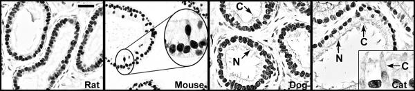
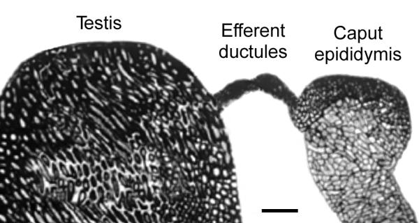
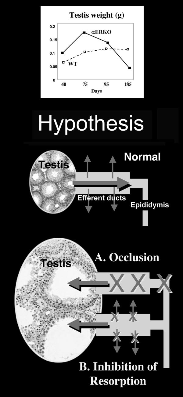
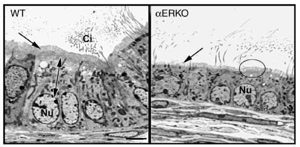
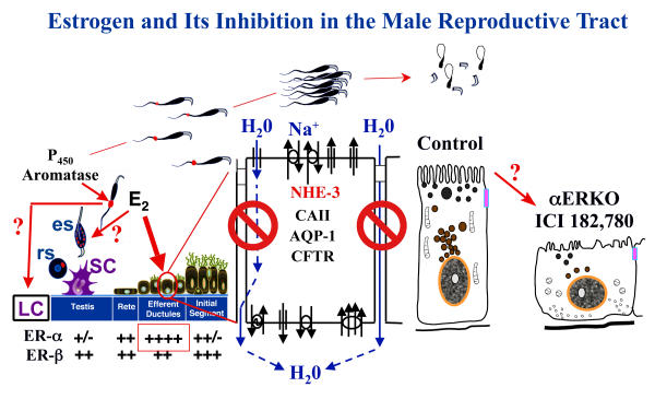
Testosterone and estrogen are no longer considered male only and female only hormones. Both hormones are important in both sexes. It was known as early as the 1930's that developmental exposure to a high dose of estrogen causes malformation of the male reproductive tract, but the early formative years of reproductive biology as a discipline did not recognize the importance of estrogen in regulating the normal function of the adult male reproductive tract. In the adult testis, estrogen is synthesized by Leydig cells and the germ cells, producing a relatively high concentration in rete testis fluid. Estrogen receptors are present in the testis, efferent ductules and epididymis of most species. However, estrogen receptor-alpha is reported absent in the testis of a few species, including man. Estrogen receptors are abundant in the efferent ductule epithelium, where their primary function is to regulate the expression of proteins involved in fluid reabsorption. Disruption of the alpha-receptor, either in the knockout (alphaERKO) or by treatment with a pure antiestrogen, results in dilution of cauda epididymal sperm, disruption of sperm morphology, inhibition of sodium transport and subsequent water reabsorption, increased secretion of Cl-, and eventual decreased fertility. In addition to this primary regulation of luminal fluid and ion transport, estrogen is also responsible for maintaining a differentiated epithelial morphology. Thus, we conclude that estrogen or its alpha-receptor is an absolute necessity for fertility in the male.


Figures

Figure 1
Figure 1
Estrogen in rete testis fluid. Mean concentrations (pg/ml) for estradiol or total estrogens in four species, rat [41], monkey [44], bull [42] and boar [46].
Figure 2
Figure 2
Estrogen sources and targets in the male reproductive tract. Estradiol 17β (E2) is produced in peripheral tissues and delivered via the plasma, but is also synthesized by Leydig cells (LC) in the testicular interstitium. The contribution of E2 from testis to plasma and from the vasculature to the testis is unknown, but it is assumed that most of the lymphatic E2 would be derived from LC. LC and germ cells (GC) contain p450 aromatase in the adult testis. LC may also contribute to the E2 concentrations in the rete testis fluid, but it is more likely that germ cell production of E2 provides the estrogen that will target the efferent ductule epithelium, the region that contains the highest concentration of ER. Less is known of E2 function and targets in the epididymis and vas deferens.
Figure 3
Figure 3
Estrogen receptor-α immunohistochemistry in the efferent ductules. ERα is abundant in the ductules of most species examined. Represented here are ductules from the rat, mouse, dog and cat [109,110,130]. Ciliated (C) and nonciliated (N) cells are strongly positive in all these species, except the cat, where ciliated cells show weak staining. Bar = 25 μm.
Figure 4
Figure 4
Testis, efferent ductules and epididymis. The surrounding fat pad was dissected away to show the efferent ductules that lie between the testis and caput epididymis. Bar = 2 mm.
Figure 5
Figure 5
Hypothesis to account for testicular weight increase in the αERKO mouse. The αERKO mouse testis was shown to increase in weight from day 40 to 75 days of age, and then the weight declined until the testis was atrophied by day 185 [59]. Two hypotheses were proposed to account for mechanisms that could explain the transient increase in testis weight prior to regression. In the normal testis, efferent ductules receive low concentrations of sperm from the rete testis. Approximately 95% of this fluid is reabsorbed by the efferent ductule epithelium, which increases the concentration of sperm that enter the epididymis. Disruption of ERα causes testicular swelling through one of two possible mechanisms: A. the efferent ductules become occluded, or B. the fluid reabsorption pathways are inhibited. Both mechanisms will result in fluid accumulation in the seminiferous tubules and backing up of fluids into the testis. Atrophy occurs by an unknown mechanism that inhibits spermatogenesis.
Figure 6
Figure 6
Histology of the efferent ductule epithelium in αERKO mouse. The wild-type (WT) ductule epithelium is columnar in shape with nonciliated cells that contain large spherical to oblong shaped nuclei (Nu) and extensive apical cytoplasm (double arrow). The nonciliated cell has a tall microvillus brush border (arrow) and extensive endocytotic apparatus. The ciliated cells have motile cilia (Ci) that extend into the lumen. The αERKO efferent ductule epithelium has a low cuboidal shape, with the apical cytoplasm reduced in size and the nucleus (Nu) also smaller. Microvilli are sparse on some cells (arrow) and reduced in height in other cells (circle). Bar = 10 μm.
Figure 7
Figure 7
Estrogen and its inhibition in the male reproductive tract: a summary. In adult males, germ cells, as well as Leydig cells (LC) contain P450 aromatase and actively synthesize estrogen (E2), which produces a relatively high concentration in rete testis fluid. This luminal estrogen targets estrogen receptors that are abundant throughout the male reproductive tract, but particularly ERα that is localized in the efferent ductule epithelium, where its expression is more abundant than even the female reproductive tract. In the testis, E2 may also feedback to influence the function of LC and spermatids, either round spermatids (rs) or elongated spermatids (es). Estrogen's primary function in the male tract is the regulation of fluid reabsorption in the efferent ductules via ERα, which increases the concentration of sperm prior to entering the epididymis. Disruption of ERα, either in the knockout (αERKO) or by treatment with a pure antiestrogen ICI 182,780, results in a decrease in Na+ transport from lumen to interstitium and thus a decrease in water (H2O) and fluid reabsorption. This inhibition is mediated by a decrease in the expression of NHE3 mRNA and protein and also decreases in carbonic anhydrase II (CAII) and aquaporin I (AQP-1) proteins. There is also an increase in cystic fibrosis transmembrane conductance regulator protein and mRNA, which adds to the NHE3 effect by secreting Cl- into the lumen by the cystic fibrosis transmembrane conductance regulator (CFTR) [64]. This inhibition of fluid reabsorption results in the dilution of cauda epididymal sperm, disruption of sperm morphology, and eventual decreased fertility. In addition to this primary regulation of luminal fluids and ions, estrogen is also responsible for maintaining a differentiated epithelial morphology through an unknown mechanism.

Similar articles

Cited by

References

    1. Wolff E, Ginglinger A. Sur la transformation des Poulets males en intersexues par injection d'hormone femelle (folliculine) aux embryons. Archs Anat Histol Embryol. 1935;20:219–278.
    1. Weniger JP. Aromatase activity in fetal gonads of mammals. J Dev Physiol. 1990;14:303–306. - PubMed
    1. Burrows H. Pathological conditions induced by oestrogenic compounds in the coagulating gland and prostate of the mouse. Am J Cancer. 1935;23:490–512.
    1. Greene RR, Burrill MW, Ivy AC. Experimental intersexuality. Am J Anat. 1940;67:305–345.
    1. Arai Y, Mori T, Suzuki Y, Bern H. Long-term effects of perinatal exposure to sex steroids and diethylstilbestrol on the reproductive system of male mammals. Int Rev Cytol. 1983;84:235–265. - PubMed

Publication types

LinkOut - more resources