

Identification of swine hepatitis E virus (HEV) and prevalence of anti-HEV antibodies in swine and human populations in Korea
- PMID: 12904362
- PMCID: PMC179837
- DOI: 10.1128/JCM.41.8.3602-3608.2003
Identification of swine hepatitis E virus (HEV) and prevalence of anti-HEV antibodies in swine and human populations in Korea
Abstract
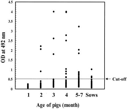
The swine hepatitis E virus (HEV) is considered to be a new zoonotic agent due to its close genomic resemblance to the human HEV and its ability to infect nonhuman primates. Hepatitis caused by HEV infection has been a serious public health problem in developing countries. However, recent seroprevalence studies indicate that the HEV also circulates in industrialized countries. In this study, a nested reverse transcription (RT)-PCR was developed to detect a part of the swine HEV open reading frame 2. Three Korean isolates of swine HEV were identified in 128 swine sera (2.3% prevalence) by the nested RT-PCR method. They were isolated from 2- to 3-month old pigs showing an age-specific prevalence of the HEV viremia. A phylogenetic tree analysis with a number of swine and human HEV isolates indicated that all Korean isolates of the swine HEV belong to genotype III. They were closely related to the swine and human HEV isolates that were identified in the United States and Japan. In addition, they formed a distinct branch in genotype III, showing a 92.7 to 99.8% identity at their nucleotide sequences. The overall prevalence of anti-swine HEV antibodies in swine was 15%. Antibodies to the swine HEV were not detected in 1-month-old pigs. However, the anti-swine HEV antibodies appeared in pigs older than 1 month and also showed an age-specific prevalence. The antibody prevalence rates to the swine HEV were 6.0, 10.0, 36.0, and 25.0%, in 2-, 3-, 4-, and 5-to-7-month-old pigs, respectively. In addition, the seroprevalence in sows to the swine HEV was 8.8%. On the other hand, 18% of blood donors in Korea were found to be positive for anti-HEV antibodies. Overall, this study indicates that subclinical HEV infections may prevail in swine and human populations in Korea.
Figures


References
-
- Aggarwal, R., and K. Krawczynski. 2000. Hepatitis E: an overview and recent advances in clinical and laboratory research. J. Gastroenterol. Hepatol. 15:9-20. - PubMed
-
- Arankalle, V. A., M. K. Goverdhan, and K. Banerjee. 1994. Antibodies against hepatitis E virus in Old World monkeys. J. Viral. Hepat. 1:125-129. - PubMed
-
- Arankalle, V. A., S. A. Tsarev, M. S. Chadha, D. W. Alling, S. U. Emerson, K. Banerjee, and R. H. Purcell. 1995. Age-specific prevalence of antibodies to hepatitis A and E viruses in Pune, India, 1982 and 1992. J. Infect. Dis. 171:447-450. - PubMed
-
- Arankalle, V. A., S. Paranjape, S. U. Emerson, R. H. Purcell, and A. M. Walimbe. 1999. Phylogenetic analysis of hepatitis E virus isolates from India (1976-1993). J. Gen. Virol. 80:1691-1700. - PubMed
Publication types
MeSH terms
Substances
LinkOut - more resources
Full Text Sources
Other Literature Sources
