

Genomic approach to identification of Mycobacterium bovis diagnostic antigens in cattle
- PMID: 12904381
- PMCID: PMC179839
- DOI: 10.1128/JCM.41.8.3719-3728.2003
Genomic approach to identification of Mycobacterium bovis diagnostic antigens in cattle
Abstract
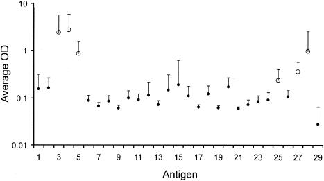
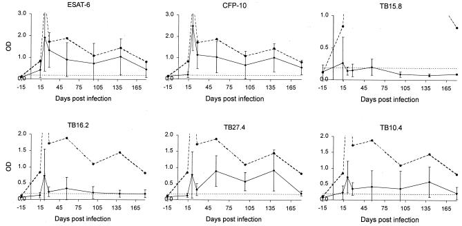
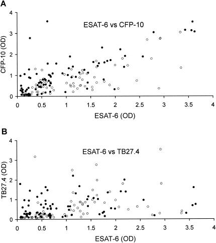
Differential delayed-type hypersensitivity skin testing with tuberculin purified protein derivatives from Mycobacterium bovis and M. avium is the standard for diagnosing bovine tuberculosis. However, improved tests based on defined, specific antigens are urgently needed. In the present study, a combination of bioinformatics, molecular biology, and bovine models of infection were used to screen mycobacterial proteins for their potential as diagnostic reagents which could be used in a whole-blood assay for diagnosis of tuberculosis. Initial screening of 28 proteins selected in silico and expressed as recombinants in Escherichia coli indicated that CFP-10, ESAT-6, TB27.4, TB16.2, TB15.8, and TB10.4 induced strong gamma interferon responses in experimentally infected cattle. A more thorough investigation over time in two groups of animals infected with a high (10(6) CFU) and a low (10(4) CFU) dose of M. bovis revealed that, for both groups, the strength of the in vitro response to individual antigens varied greatly over time. However, combining the results for ESAT-6, CFP-10, and TB27.4, possibly supplemented with TB10.4, gave sensitivities at different infection stages close to those obtained with M. bovis purified protein derivative. Importantly, while responsiveness to ESAT-6 and CFP-10 correlated strongly for individual samples, the same was not the case for ESAT-6 and TB27.4 responsiveness. The results suggest that combinations of specific antigens such as these have great potential in development of optimized diagnostic systems for bovine tuberculosis.
Figures
References
-
- Amadori, M., S. Tagliabue, S. Lauzi, G. Finazzi, G. Lombardi, P. Telò, L. Pacciarini, and L. Bonizzi. 2002. Diagnosis of Mycobacterium bovis infection in calves sensitized by mycobacteria of the avium/intracellulare group. J. Vet. Med. B Infect. Dis. Vet. Public Health 49:89-96. - PubMed
-
- Andersen, P., M. E. Munk, J. M. Pollock, and T. M. Doherty. 2000. Specific immune-based diagnosis of tuberculosis. Lancet 356:1099-1104. - PubMed
-
- Arend, S. M., A. C. Engelhard, G. Groot, K. de Boer, P. Andersen, T. H. Ottenhoff, and J. T. van Dissel. 2001. Tuberculin skin testing compared with T-cell responses to Mycobacterium tuberculosis-specific and nonspecific antigens for detection of latent infection in persons with recent tuberculosis contact. Clin. Diagn. Lab. Immunol. 8:1089-1096. - PMC - PubMed
-
- Behr, M. A., M. A. Wilson, W. P. Gill, H. Salamon, G. K. Schoolnik, S. Rane, and P. M. Small. 1999. Comparative genomics of BCG vaccines by whole-genome DNA microarray. Science 284:1520-1523. - PubMed
-
- Berthet, F.-X., P. B. Rasmussen, I. Rosenkrands, P. Andersen, and B. Gicquel. 1998. A Mycobacterium tuberculosis operon encoding ESAT-6 and a novel low-molecular-mass culture filtrate protein (CFP-10). Microbiology 144:3195-3203. - PubMed
Publication types
MeSH terms
Substances
LinkOut - more resources
Full Text Sources
Miscellaneous
