

Evidence of parvovirus replication in cerebral neurons of cats
- PMID: 12904392
- PMCID: PMC179857
- DOI: 10.1128/JCM.41.8.3801-3805.2003
Evidence of parvovirus replication in cerebral neurons of cats
Abstract
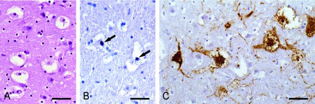
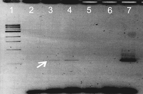
The correlation between parvovirus infections and lesions in the central nervous system other than cerebellar hypoplasia was studied in 100 cats. The animals were necropsied with a history of various diseases, one third showing typical clinical and pathomorphological signs of panleukopenia. In 18 cats polyclonal antiserum against canine parvovirus consistently labeled neurons mainly in diencephalic regions, whereas the cerebellar cortex remained negative in all cases. In situ hybridization with digoxigenin-labeled minus-sense RNA probes, hybridizing with monomer-replicative form DNA or mRNA, revealed positive signals in nuclei of several neurons of the brain, again excluding the cerebellum. PCR applied to formalin-fixed and paraffin-embedded brain tissue and intestinal tissues of the diseased cats and subsequent DNA sequence analysis yielded canine parvovirus type 2 (CPV-2)-like sequences in the central nervous system. Two aspects of these findings are intriguing: (i). parvoviruses appear to be capable of replicating in neurons, cells that are considered to be terminally differentiated and (ii). CPV-like viruses of the old antigenic type CPV-2 appear to be able to infect cats.
Figures
References
-
- Csiza, C. K., A. de Lahunta, F. W. Scott, and J. H. Gillespie. 1972. Spontaneous feline ataxia. Cornell Vet. 62:300-322. - PubMed
-
- Csiza, C. K., F. W. Scott, A. de Lahunta, and J. H. Gillespie. 1972. Respiratory signs and central nervous system lesions in cats infected with panleukopenia virus. A case report. Cornell Vet. 62:192-195. - PubMed
-
- Hervàs, G., J. Calvo Marqués, and M. Pumarola Batlle. 1999. Cerebellar hypoplasia in the cat. Eur. J. Comp. Anim. Pract. 9:47-52.
MeSH terms
Substances
LinkOut - more resources
Full Text Sources
Miscellaneous
