. 2003 Aug 6;23(18):7069-74.
doi: 10.1523/JNEUROSCI.23-18-07069.2003.

Selective antagonism of GluR5 kainate-receptor-mediated synaptic currents by topiramate in rat basolateral amygdala neurons

Affiliations

Selective antagonism of GluR5 kainate-receptor-mediated synaptic currents by topiramate in rat basolateral amygdala neurons

Divina S Gryder et al. J Neurosci. .

Abstract

Topiramate is a widely used antiepileptic agent whose mechanism of action is poorly understood. The drug has been reported to interact with various ion channel types, including AMPA/kainate receptors. In whole-cell voltage-clamp recordings from principal neurons of the rat basolateral amygdala, topiramate at low concentrations (IC50, approximately 0.5 microm) selectively inhibited pharmacologically isolated excitatory synaptic currents mediated by kainate receptors containing the GluR5 subunit. Topiramate also partially depressed predominantly AMPA-receptor-mediated EPSCs, but with lower efficacy. Topiramate did not alter the degree of facilitation in paired-pulse experiments, and it reduced the amplitude of miniature EPSCs without affecting their frequency, demonstrating that the block of synaptic responses occurs postsynaptically. Inhibition of GluR5 kainate receptors could represent a key mechanism underlying the anticonvulsant activity of topiramate. Moreover, these results support the concept that GluR5 kainate receptors represent a novel target for antiepileptic drug development.


Figures
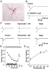
Figure 1.
Figure 1.
LY 293558 and topiramate block GluR5 kainate-receptor-mediated synaptic responses in BLA neurons. A, Typical biocytin-filled neuron in a slice stained for horseradish peroxidase and counterstained with cresyl violet. Scale bar, 100 μm. B, Synaptic responses were evoked by EC stimulation (triangles) in the presence of AP5, GYKI 52466, bicuculline, and SCH 50911. Perfusion for 10 min with 10 μm LY 293558 eliminated the residual synaptic response representing the component mediated by GluR5 kainate receptors. Holding potential, -60 mV. C, Depression of GluR5 kainate receptor responses by 1 nm (top) and 1 μm (bottom) topiramate. The topiramate traces were obtained 40 min after the onset of the topiramate perfusion; the wash traces were obtained 8-12 min after the termination of the topiramate perfusion. Each trace is the average of the responses to 25 stimuli. D, Time course of the development of inhibition in the experiment with 1 μm topiramate. Data points indicate mean peak amplitude values for 25 successive stimuli applied at 10 sec intervals normalized to the mean peak value before topiramate (SE indicated by gray band). E, Concentration-response curve for the inhibition of GluR5 kainate receptor current by various concentrations of topiramate. Each point represents the mean ± SEM of the steady-state level of inhibition 20 - 60 min after the onset of topiramate perfusion in two to four experiments.
Figure 2.
Figure 2.
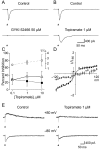
A, Block of predominantly AMPA-receptor-mediated synaptic current by 50 μm GYKI 52466. The recording was performed in the presence of AP5, LY 293558, bicuculline, and SCH 50911. B, Partial inhibition of the predominantly AMPA-receptor-mediated synaptic current by 1 μm topiramate. Note the prolongation of the time course of the response in the presence of topiramate. In A and B, each trace is the average of the responses to 25 stimuli. C, Concentration-response curves for topiramate inhibition of AMPA-receptor-mediated synaptic currents. Current decay in the presence of topiramate was fitted by a single exponential function; the fits for the responses to 25 successive stimuli were averaged. The percent inhibition of the peak current values (filled squares) and the zero time values from the exponential fits (open triangles) are plotted with respect to the left ordinate. The ratios of the decay time constant values from fits in the presence (τ) and absence (τc) of topiramate are plotted with respect to the right ordinate. Each point represents the mean ± SEM of values from two to four experiments. D, Voltage dependence of evoked GluR5 kainate-receptor-mediated synaptic currents. The peak amplitude of synaptic responses in the absence (filled squares) and presence (open squares) of 1 μm topiramate was determined at various holding potential levels and normalized to the control value for that cell at -60 mV (I-60). Each point represents the mean ± SEM of three control values and seven topiramate values from three cells. The fits are arbitrary. E, Sample traces comparing the inhibitory action of topiramate on evoked GluR5 kainate receptor currents at -80 and +80 mV.
Figure 3.
Figure 3.
Effect of topiramate on paired-pulse facilitation of GluR5 kainate-receptor-mediated evoked synaptic currents. A, Perfusion with 10 μm topiramate substantially reduces the amplitude of both responses (40 min after onset of perfusion) with partial recovery during wash (12 min after termination of topiramate). Triangles indicate paired-pulse stimulation. B, Superposition of control, topiramate, and wash traces from A. C, Summary of data from four separate experiments similar to that shown in A. Each bar represents the ratio (P2 /P1) of the peak amplitudes of the synaptic responses to the second stimulus and the first stimulus. There is no significant difference between the control, topiramate, and wash values.
Figure 4.
Figure 4.
Effect of topiramate on spontaneous GluR5 kainate-receptor-mediated mEPSCs in a BLA neuron. The recording was performed in the presence of 100 μm AP5, 50 μm GYKI 52466, 10 μm bicuculline, 10 μm SCH 50911, and 1 μm tetrodotoxin. A, Twenty representative aligned mEPSCs; the white trace is the average. B, Averages of 20 mEPSCs before and 15 min after onset of 10 μm topiramate perfusion, and immediately after the termination of the perfusion (wash). C, mEPSCs during 15 min epochs were collected and the area (representing the charge) was determined by integration over a 60 msec period. The histogram plots the normalized integrated charge values for mEPSCs collected under the conditions as in B. mEPSCs were detected in a nonbiased manner using a scaled template procedure based on an average of five control mEPSCs. Mean ± SE rise and decay time constants were control: 7.2 ± 0.2 and 23.6 ± 0.8 (n = 480); topiramate: 4.9 ± 0.2 and 16.5 ± 0.4 (n = 561); wash: 8.2 ± 0.7 and 34.2 ± 3.4 (n = 73). Gaussian fits to the control, topiramate, and wash histograms gave center values of 56.0, 4.8, and 33.4 fC. Similar results were obtained in five additional experiments.

References

    1. Bleakman D, Lodge D ( 1998) Neuropharmacology of AMPA and kainate receptors. Neuropharmacology 37: 1187-1204. - PubMed
    1. Bleakman D, Gates MR, Ogden AM, Mackowiak M ( 2002) Kainate receptor agonists, antagonists and allosteric modulators. Curr Pharm Des 8: 873- 885. - PubMed
    1. Burnashev N, Villarroel A, Sakmann B ( 1996) Dimensions and ion selectivity of recombinant AMPA and kainate receptor channels and their dependence on Q/R site residues. J Physiol (Lond) 496 (Pt 1): 165-173. - PMC - PubMed
    1. Castillo PE, Malenka RC, Nicoll RA ( 1997) Kainate receptors mediate a slow postsynaptic current in hippocampal CA3 neurons. Nature 388: 182-186. - PubMed
    1. Chong MS, Libretto SE ( 2003) The rationale and use of topiramate for treating neuropathic pain. Clin J Pain 19: 59-68. - PubMed

MeSH terms

LinkOut - more resources