. 2003 Aug 6;23(18):7084-92.
doi: 10.1523/JNEUROSCI.23-18-07084.2003.

Insulin receptor substrate-2 deficiency impairs brain growth and promotes tau phosphorylation

Affiliations

Insulin receptor substrate-2 deficiency impairs brain growth and promotes tau phosphorylation

Markus Schubert et al. J Neurosci. .

Abstract

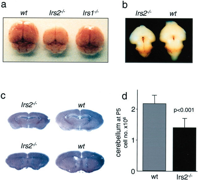
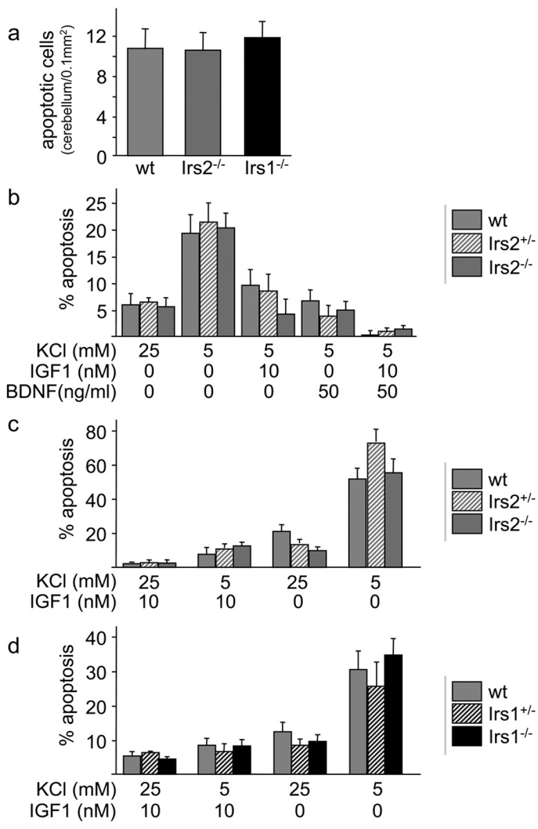
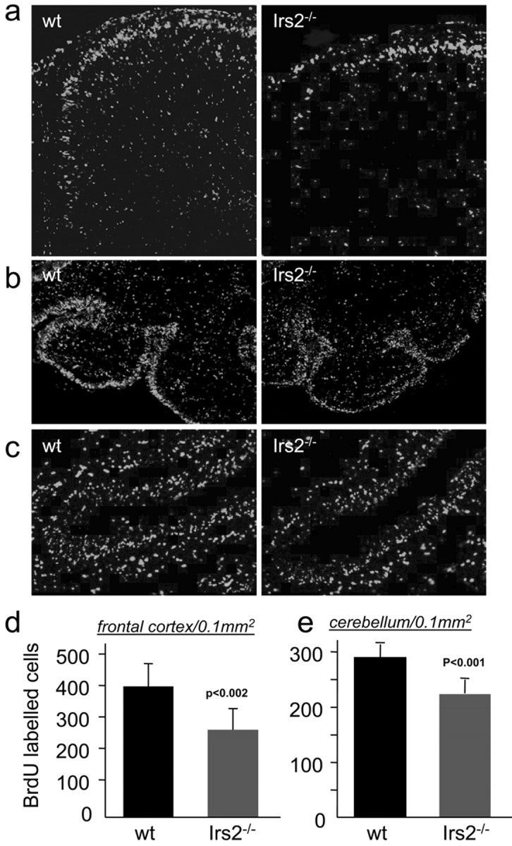
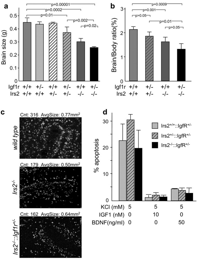
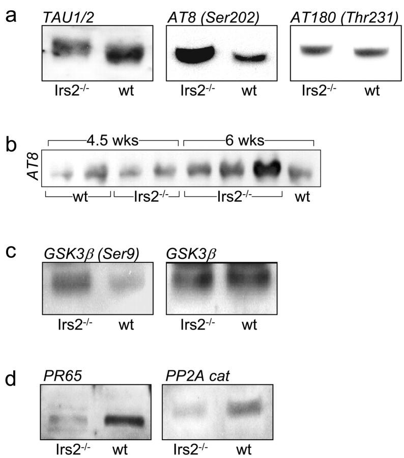
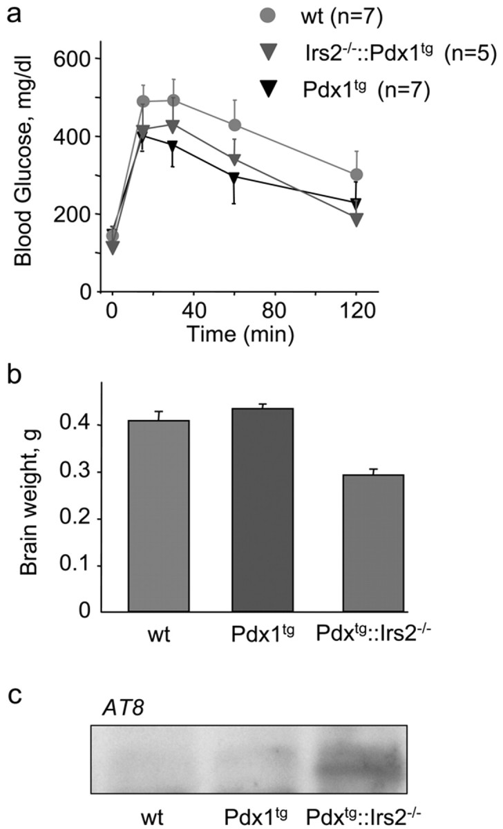
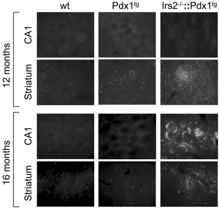
Insulin resistance and diabetes might promote neurodegenerative disease, but a molecular link between these disorders is unknown. Many factors are responsible for brain growth, patterning, and survival, including the insulin-insulin-like growth factor (IGF)-signaling cascades that are mediated by tyrosine phosphorylation of insulin receptor substrate (IRS) proteins. Irs2 signaling mediates peripheral insulin action and pancreatic beta-cell function, and its failure causes diabetes in mice. In this study, we reveal two important roles for Irs2 signaling in the mouse brain. First, disruption of the Irs2 gene reduced neuronal proliferation during development by 50%, which dissociated brain growth from Irs1-dependent body growth. Second, neurofibrillary tangles containing phosphorylated tau accumulated in the hippocampus of old Irs2 knock-out mice, suggesting that Irs2 signaling is neuroprotective. Thus, dysregulation of the Irs2 branch of the insulin-Igf-signaling cascade reveals a molecular link between diabetes and neurodegenerative disease.


Figures

Figure 1.
Figure 1.
Brains of Irs2-/- mice are reduced in size. a, Brains of Irs2-/-, Irs1-/-, and wild-type (wt) mice at 4 weeks of age. b, Brains of Irs2-/- and wild-type mice at E16. Embryos were collected from timed pregnant mothers and fixed in 4% paraformaldehyde. All visible brain regions of the Irs2-/- appear small but normal. c, HE-stained sections of Irs2-/- and wild-type brains showing a global reduction in all regions. d, Total cell number of the cerebella of Irs2-/- and wild-type mice at P5. Cerebella were dispersed and the total cell number was determined (n = 6 per group).
Figure 2.
Figure 2.
Apoptosis in brains and cultured neurons from mice lacking IRS proteins. a, TUNEL assay of the cerebellum at P6 reveals no differences between Irs2-/-, Irs1-/-, and wild-type (wt) mice. Quantification of apoptotic nuclei showed the same number per area in all genotypes. b, IGF1 and BDNF protect against neuronal apoptosis in the absence of Irs2. Cerebellar granule cells (P5) were cultured for 4 d in medium containing normal serum. Neurons were then incubated for an additional 24 hr in serum-free media containing 5 mm KCl, 5 mm KCl plus 10 nm IGF1, 5 mm KCl plus 50 ng/ml BDNF, or 25 mm KCL. Cells were stained with Hoechst 33342 and counted. Percentage apoptosis was calculated (n = 3 per genotype and experiment; 3 independent experiments showed similar results). c, Cerebellar granule cells from Irs2+/- × Irs2+/- litters (Irs2+/- or Irs2-/- cells) were cultured for 4 d, followed by incubation for 24 hr in serum-free media containing 25 mm KCl plus 10 nm IGF1 (+KCl, +IGF1), 10 nm IGF1 (-KCl, +IGF1), 25 mm KCl (+KCl, -IGF1), or minimal medium (-KCl, -IGF1) (n = 2 per genotype and experiment; 3 independent experiments showed similar results). d, Cerebellar granule cells from Irs1+/- × Irs1+/- litters (Irs1+/- or Irs1-/- cells) were cultured for 4 d, followed by incubation for 24 hr as described in c.
Figure 3.
Figure 3.
Neuronal proliferation is impaired in Irs2-/- brains. a, Four pregnant mice were injected intraperitoneally with BrdU at E14, and the embryos were harvested after 48 hr. Serial sections of the frontal cortex were obtained, and the total number of BrdU-labeled neurons per area was quantified using the Openlab software (Improvision). b, Mice (P6) were injected with BrdU, brains were fixed after 24 hr, and cryostat sections of the cerebellum were obtained (n = 4 per group). Sections were stained using an anti-BrdU antibody (Boehringer Mannheim). Labeled neurons were counted using the Openlab software (Improvision). c, Enlargement of b, demonstrating labeled neurons in the outer and inner granule layer. d,e, Quantification of BrdU-labeled cells in the frontal cortex (d) and cerebellum (e) from wild-type (wt) or Irs2-/- sections.
Figure 4.
Figure 4.
Igf1r heterozygosity causes a further reduction in brain size in Irs2-/- mice. a, Brain size of 6-week-old wild-type (wt), Igf1r+/-, Irs2+/-, Irs2+/-Igf1r+/-, Irs2-/-, and Igf1r+/-::Irs2-/- mice. b, Brain-body ratio (%) of 6-week-old mice of the different genotypes. c, BrdUincorporation in cerebella granule cells at P6 (48 hr survival) from wild type, Irs2-/-, or Irs2-/-Igf1r+/-. Cell counts and average cell size were calculated with NIH ImageJ. d, Apoptosis in the cerebella granule cells from Igf1r+/-, Irs2+/-Igf1r+/-, and Irs2-/-Igf1r+/- mice (P5). Granule cells were cultured for 4 d and then incubated for an additional 24 hr in serum-free medium containing 5 mm KCl,5mm KCl plus 10 nm IGF1,5 mm KCl plus 50 ng/ml BDNF. pvalues were calculated using Sigmaplot (Sigma, St. Louis, MO) version 8.1.
Figure 5.
Figure 5.
Immunoblotting with various antibodies conducted with whole-brain lysates. a, Lysates from 6-week-old mice were immunoblotted with antibodies against tau1/2 protein, the AT8 antibody against tau phosphorylated at Ser202, and the AT180 antibody against tau phosphorylated at Thr231. b, Lysates from 4.5- and 6-week-old mice were immunoblotted with AT8 antibody against tau phosphorylated at Ser202. c, Lysates from 6-week-old mice were immunoblotted with antibodies against phospho-GSK3β (Ser9) and GSK3β protein. d, Lysates from 6-week-old mice were immunoblotted with antibodies against PP2A scaffold subunit (PR65) and PP2A catalytic subunits. wt, Wild type.
Figure 6.
Figure 6.
Glucose tolerance, brain size, and tau phosphorylation in wild-type (wt), Pdx1tg, and Irs2-/-::Pdx1tg mice. a, Glucose tolerance was measured at 14 months of age. b, Brain weight at ∼14 months of age. c, Western blot analysis using phospho-tau Ser202 antibody (AT8) of whole-brain lysates at 16 months of age.
Figure 7.
Figure 7.
AT8 immunostaining from 12- or 16-month-old brains of wild-type (wt), Pdx1tg, and Irs2-/-::Pdx1tg mice. Pyramidal cells of the CA1 region or coronal sections through the corpus striatum (Striatum) were stained with the AT8 antibody (CA1; original magnification, 200×).

References

    1. Aberg M, Aberg N, Hedbacker H, Oscarsson J, Eriksson P ( 2000) Peripheral infusion of IGF-1 selectively induces neurogenesis in the adult rat hippocampus. J Neurosci 20: 2896-2903. - PMC - PubMed
    1. Aguirre V, Werner ED, Giraud J, Lee YH, Shoelson SE, White MF ( 2002) Phosphorylation of ser307 in insulin receptor substrate-1 blocks interactions with the insulin receptor and inhibits insulin action. J Biol Chem 277: 1531-1537. - PubMed
    1. Airey DC, Lu L, Williams RW ( 2001) Genetic control of the mouse cerebellum: identification of quantitative trait loci modulating size and architecture. J Neurosci 21: 5099-5109. - PMC - PubMed
    1. Anlar B, Sullivan KA, Feldman EL ( 1999) Insulin-like growth factor-I and central nervous system development. Horm Metab Res 31: 120-125. - PubMed
    1. Bektas A, Warram JH, White MF, Krolewski AS, Doria A ( 1999) Exclusion of insulin receptor substrate 2 (IRS-2) as a major locus for early-onset autosomal dominant type 2 diabetes. Diabetes 48: 640-642. - PubMed

Publication types

MeSH terms