. 2003 Aug 6;23(18):7093-106.
doi: 10.1523/JNEUROSCI.23-18-07093.2003.

A broad role for melanopsin in nonvisual photoreception

Affiliations

A broad role for melanopsin in nonvisual photoreception

Joshua J Gooley et al. J Neurosci. .

Abstract

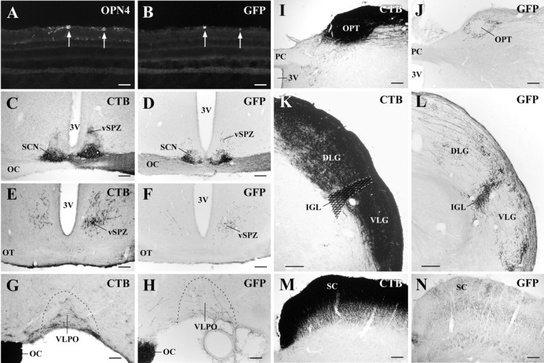
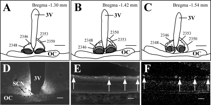
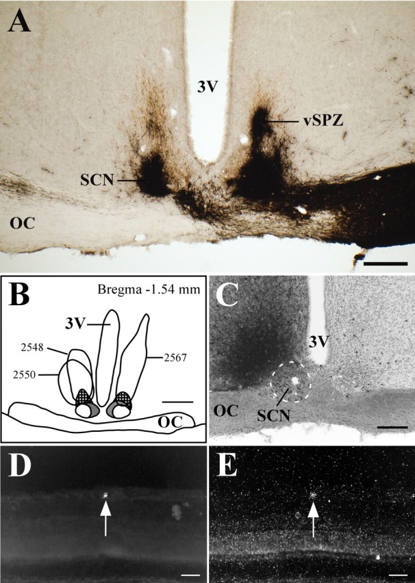
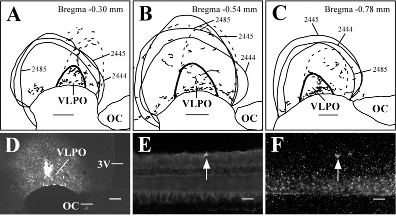
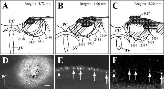
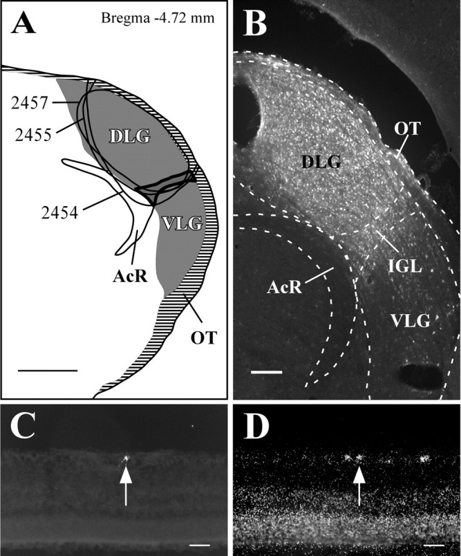
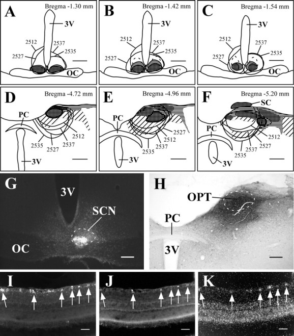
The rod and cone photoreceptors that mediate visual phototransduction in mammals are not required for light-induced circadian entrainment, negative masking of locomotor activity, suppression of pineal melatonin, or the pupillary light reflex. The photopigment melanopsin has recently been identified in intrinsically photosensitive retinal ganglion cells (RGCs) that project to the suprachiasmatic nucleus (SCN), intergeniculate leaflet (IGL), and olivary pretectal nucleus, suggesting that melanopsin might influence a variety of irradiance-driven responses. We have found novel projections from RGCs that express melanopsin mRNA to the ventral subparaventricular zone (vSPZ), a region involved in circadian regulation and negative masking, and the sleep-active ventrolateral preoptic nucleus (VLPO) and determined the subsets of melanopsin-expressing RGCs that project to the SCN, the pretectal area (PTA), and the IGL division of the lateral geniculate nucleus (LGN). Melanopsin was expressed in the majority of RGCs that project to the SCN, vSPZ, and VLPO and in a subpopulation of RGCs that innervate the PTA and the IGL but not in RGCs projecting to the dorsal LGN or superior colliculus. Two-thirds of RGCs containing melanopsin transcript projected to each of the SCN and contralateral PTA, and one-fifth projected to the ipsilateral IGL. Double-retrograde tracing from the SCN and PTA demonstrated a subpopulation of RGCs projecting to both sites, most of which contained melanopsin mRNA. Our results suggest that melanopsin expression defines a subset of RGCs that play a broad role in the regulation of nonvisual photoreception, providing collateralized projections that contribute to circadian entrainment, negative masking, the regulation of sleep-wake states, and the pupillary light reflex.


Figures

Figure 1.
Figure 1.
Anterograde tracing from RGCs transduced by rAAV-GFP demonstrates primarily input from melanopsin-expressing RGCs. A, B, D, F, H, J, L, N, Case 2472 after intravitreal injection with rAAV-GFP. C, E, G, I, K, M, Case 1172 after intravitreal injection with CTB. After injection of rAAV-GFP into the vitreous body of the right eye, 81% of GFP-producing cells in the ganglion cell layer (B) also contained melanopsin (A). Arrows indicate double-labeled cells. GFP-immunoreactive axonal terminals were observed in the SCN (D), vSPZ (F), VLPO (H), OPT (J), and IGL (L). In contrast, GFP-labeled projections to the DLG and VLG nuclei were much less intense than those to the IGL. The bilateral SCN and vSPZ and the contralateral VLPO, OPT, IGL, and SC are shown. 3V, Third ventricle; OC, optic chiasm; OPN4, melanopsin; OT, optic tract; PC, posterior commissure.Scale bars: A, B, 50 μm; C-F, I-N, 200 μm; G-H, 100 μm.
Figure 2.
Figure 2.
Melanopsin is expressed in the majority of RGCs that project to the SCN. A-C, Camera lucida drawings of coronal brain sections from FG-injected animals. The ventrolateral SCN is checkered, the dorsomedial SCN is colored gray, and the dashed outline dorsal to the SCN indicates the part of the vSPZ that receives relatively sparse retinal input. Smoothly drawn lines indicate injection sites. All injections were made in the right SCN, but some are transposed to the left side for clarity. D-F, Case 2348. Injections of FG in the SCN (D) resulted in retrogradely labeled RGCs in the contralateral eye (E) that were also positive for melanopsin transcript (F). Arrows indicate double-labeled cells. 3V, Third ventricle; OC, optic chiasm. Scale bars: A-C, 500 μm; D, 200 μm; E, F, 50 μm.
Figure 3.
Figure 3.
Melanopsin is expressed in the majority of RGCs that project to the vSPZ. A, Retinal terminals (stained in black) overlap extensively with the contralateral vSPZ, as defined by the column of VIP-immuoreactive fibers (stained in brown) leaving the dorsal margin of the SCN. B, Camera lucida drawing of a coronal brain section from BDA-CTB-injected animals. The checkered region indicates the retinorecipient vSPZ, and the gray and white regions ventral to the vSPZ represent the dorsomedial and ventrolateral SCN, respectively. Smoothly drawn lines indicate injection sites. All injections were made in the right vSPZ, but some are transposed to the left side for clarity. C-E, Case 2550. Injections of BDA-CTB in the area including the vSPZ resulted in retrogradely labeled neurons throughout the ipsilateral SCN (C). Retrogradely labeled RGCs in the contralateral eye (D) were positive for melanopsin transcript (E). Arrows indicate a double-labeled cell. 3V, Third ventricle; OC, optic chiasm. Scale bars: A, C, 200 μm; B, 500 μm; D, E, 50 μm.
Figure 4.
Figure 4.
Melanopsin is expressed in the majority of RGCs that project to the VLPO. A-C, Camera lucida drawings of retinal varicosities that terminate in the VLPO region. The VLPO cluster is thickly outlined, the extended VLPO is outlined with the dashed line, and injection sites are indicated by smoothly drawn lines. D-F, Case 2445. Injections of FG in the VLPO region (D) resulted in retrogradely labeled RGCs in the contralateral eye (E) that were also positive for melanopsin transcript (F). Arrows indicate a double-labeled cell. 3V, Third ventricle; OC, optic chiasm. Scale bars: A-D, 200 μm; E, F, 50 μm.
Figure 5.
Figure 5.
Melanopsin is expressed in a subpopulation of RGCs that project to the PTA. A-C, Camera lucida drawings of coronal brain sections from FG-injected animals. The OPT is thickly outlined, and smoothly drawn lines indicate injection sites. Checkered, horizontal, and diagonal hatching indicate brain regions that are heavily, moderately, or lightly innervated by retinal efferents. D-F, Case 2438. Injections of FG in the PTA that included the OPT (D) resulted in retrogradely labeled RGCs in the contralateral eye (E) that were also positive for melanopsin transcript (F). Arrows indicate double-labeled cells. 3V, Third ventricle; PC, posterior commissure. Scale bars: A--C, 500 μm; D, 200 μm; E, F, 50 μm.
Figure 6.
Figure 6.
Melanopsin is expressed in a subpopulation of RGCs that project to the LGN. A, Camera lucida drawing of a coronal brain section from FG-injected animals. The IGL is thickly outlined, and smoothly drawn lines indicate injection sites. Gray regions indicate brain regions that receive heavy input from the retina, and horizontal lines indicate the optic tract. B-D, Case 2455. Injections of FG in the LGN that included the IGL (B) resulted in retrogradely labeled RGCs in the ipsilateral eye (C) that were also positive for melanopsin transcript (D). Arrows indicate a double-labeled cell. AcR, Acoustic radiation; OT, optic tract. Scale bars: A, 500 μm; B, 200 μm; C, D, 50 μm.
Figure 7.
Figure 7.
Melanopsin is expressed in a subset of RGCs, the axons of which bifurcate and project to both the SCN and PTA. A-F, Camera lucida drawings of coronal sections from FG- and CTB-injected animals. The retinorecipient ventrolateral SCN (A-C) is checkered, the dorsomedial SCN is colored gray, and the outlined region dorsal to the SCN indicates the sparsely innervated vSPZ. Smoothly drawn lines indicate FG injection sites. All injections were made in the left SCN, but some are transposed to the right side for clarity. The OPT is thickly outlined (D-F), and smoothly drawn lines indicate CTB injection sites. Checkered, horizontal, and diagonal hatching indicate brain regions that are heavily, moderately, or lightly innervated by retinal efferents. G-K, Case 2537. Injections of FG and CTB in the SCN (G) and PTA (H), respectively, resulted in retrogradely labeled RGCs in the contralateral eye that were both FG-immunoreactive (I) and CTB-immunoreactive (J) and were positive for melanopsin transcript (K). Arrows indicate triple-labeled cells. 3V, Third ventricle; OC, optic chiasm; PC, posterior commissure. Scale bars: A-F, 500 μm; G-H, 200 μm; I-K, 50 μm.
Figure 8.
Figure 8.
Schematic diagram showing the known projections of RGCs that express melanopsin. The density of the retinal projections is roughly indicated by thickness of the arrows. The solid branched arrow to the SCN and PTA indicates collateralized projections, and the branched dashed arrow to the SCN and IGL indicates proposed axon collaterals. Projections that might arise from melanopsin-negative RGCs to the SCN, vSPZ, VLPO, PTA, and IGL are not shown. Long dashed arrows indicate physiologic and behavioral outputs of the targeted retinorecipient brain areas. Direct projections between retinorecipient brain areas are shown, but indirect projections are not shown for reasons of clarity. Opn4+ RGCs, Melanopsin-positive RGCs; ON, optic nerve; OT, optic tract; RHT, retinohypothalamic tract. Drawing is not to scale.

References

    1. Abercrombie M ( 1946) Estimation of nuclear population from microtome sections. Anat Rec 94: 239-247. - PubMed
    1. Aggelopoulos NC, Meissl H ( 2000) Responses of neurones of the rat suprachiasmatic nucleus to retinal illumination under photopic and scotopic conditions. J Physiol (Lond) 523: 211-222. - PMC - PubMed
    1. Ali RR, Reichel MB, De Alwis M, Kanuga N, Kinnon C, Levinsky RJ, Hunt DM, Bhattacharya SS, Thrasher AJ ( 1998) Adeno-associated virus gene transfer to mouse retina. Hum Gene Ther 9: 81-86. - PubMed
    1. Auricchio A, Kobinger G, Anand V, Hildinger M, O'Connor E, Maguire AM, Wilson JM, Bennett J ( 2001) Exchange of surface proteins impacts on viral vector cellular specificity and transduction characteristics: the retina as a model. Hum Mol Genet 10: 3075-3081. - PubMed
    1. Benca RM, Overstreet DE, Gilliland MA, Russell D, Bergmann BM, Obermeyer WH ( 1996) Increased basal REM sleep but no difference in dark induction or light suppression of REM sleep in Flinders rats with cholinergic supersensitivity. Neuropsychopharmacology 15: 45-51. - PubMed

Publication types

MeSH terms

LinkOut - more resources