. 2003 Aug 19;100(17):9958-61.
doi: 10.1073/pnas.1733025100. Epub 2003 Aug 6.

Nestin expression in hair follicle sheath progenitor cells

Affiliations

Nestin expression in hair follicle sheath progenitor cells

Lingna Li et al. Proc Natl Acad Sci U S A. .

Abstract

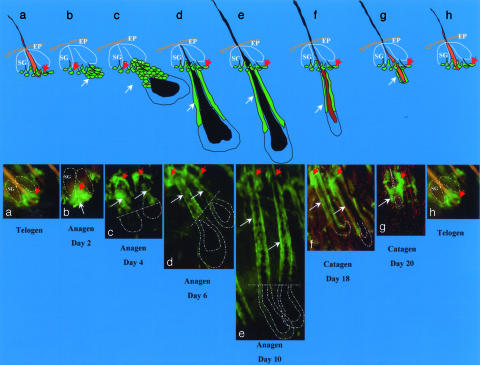
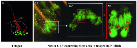
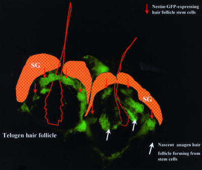
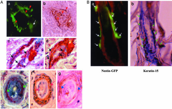
The intermediate filament protein, nestin, marks progenitor cells of the CNS. Such CNS stem cells are selectively labeled by placing GFP under the control of the nestin regulatory sequences. During early anagen or growth phase of the hair follicle, nestin-expressing cells, marked by GFP fluorescence in nestin-GFP transgenic mice, appear in the permanent upper hair follicle immediately below the sebaceous glands in the follicle bulge. This is where stem cells for the hair follicle outer-root sheath are thought to be located. The relatively small, oval-shaped, nestin-expressing cells in the bulge area surround the hair shaft and are interconnected by short dendrites. The precise locations of the nestin-expressing cells in the hair follicle vary with the hair cycle. During telogen or resting phase and in early anagen, the GFP-positive cells are mainly in the bulge area. However, in mid- and late anagen, the GFP-expressing cells are located in the upper outer-root sheath as well as in the bulge area but not in the hair matrix bulb. These observations show that the nestin-expressing cells form the outer-root sheath. Results of the immunohistochemical staining showed that nestin, GFP, keratin 5/8, and keratin 15 colocalize in the hair follicle bulge cells, outer-root sheath cells, and basal cells of the sebaceous glands. These data indicate that nestin-expressing cells, marked by GFP, in the hair follicle bulge are indeed progenitors of the follicle outer-root sheath. The expression of the unique protein, nestin, in both neural stem cells and hair follicle stem cells suggests their possible relation.


Figures

Fig. 1.
Fig. 1.
Hair follicle stem cells in the hair-growth cycle. (Upper) Cartoon showing position of nestin-GFP stem cells at each stage of the hair follicle cycle. SG, sebaceous gland; EP, epidermis. (Lower a) Nestin-GFP-expressing hair follicle stem cells (red arrow) located in the hair follicle bulge area in telogen phase. (b) Day 2 after anagen induction by depilation. Note the new hair follicle cells (white arrow) formed directly from the bulge nestin-GFP-expressing stem cells. (ce)Day4(c), day 6 (d), and day 10 (e) after anagen induction by depilation. Note the nestin-GFP-expressing outer-root sheath cells (white arrows) in the upper two-thirds of the hair follicle. (f and g) Day 19 (f) and day 20 (g) after depilation. Note in f and g that the hair follicles are in the catagen phase and are undergoing regression and degeneration, including the nestin-GFP-expressing cells in the outer-root sheath. The bulge area nestin-GFP-expressing stem cells remain. (h) Hair follicle cycling in telogen phase.
Fig. 2.
Fig. 2.
Hair follicle nestin-GFP-expressing cells in the telogen phase of nestin-GFP transgenic mouse skin. The skin sample was prepared freshly right after excision from the back skin of a nestin-GFP transgenic mouse. The skin sample then was directly observed by fluorescence or confocal microscopy with the dermis side up after s.c. tissue was dissected out. (a) Cartoon of telogen hair follicle showing position of nestin-GFP-expressing hair follicle stem cells. (a1) Low-magnification fluorescence microscopy image showing the ring of bulge nestin-GFP-expressing stem cells (small white box, see a). (a2) High-magnification confocal microscopy image reflecting the small white box in a1. Note the small round- or oval-shaped nestin-GFP-expressing cells in the bulge area of the hair follicle (small red box). (a3) High-magnification fluorescence microscopy image showing two individual nestin-GFP-expressing stem cells reflecting the red box in a2. Note the unique morphology of the hair follicle stem cells and multiple dendrite-like structures of each cell. Red arrows indicate the cell body, and red arrowheads show the multiple dendritic structure of each cell. (Original magnifications: a1, ×100; a2, ×400; a3, ×1,600.) SG, sebaceous gland; EP, epidermis.
Fig. 3.
Fig. 3.
Telogen-anagen transition showing hair follicle stem cells forming nascent hair follicle. The image was taken by fluorescence microscopy 18 h after depilation. Note the bulge nestin-GFP-expressing hair follicle stem cells in the telogen phase (left hair follicle, red arrows) and the nascent anagen hair follicle directly formed from the bulge nestin-GFP-expressing stem cells (right hair follicle, white arrows). (Original magnification, ×400.)
Fig. 4.
Fig. 4.
(A) Colocalization of GFP, nestin, and keratins 5/8 and 15 in hair follicle bulge stem cells and outer-root sheath cells determined by immunohistochemical staining. (a) Confocal image of live tissue showing nestin-GFP-expressing hair follicle bulge stem cells (red arrows) forming the nascent anagen hair follicle (white arrow). (b) Paraffin-embedded tissue section immunohistochemically stained with nestin antibody showing the colocalization of nestin expression in the nascent hair follicle and bulge stem cells. Red arrows indicate nestin-positive hair follicle bulge stem cells. The white arrow indicates nestin-positive nascent hair follicle. (c and d) Two serial paraffin-embedded longitudinal sections of nestin-GFP transgenic mouse hair follicles double-immunohistochemical-stained with GFP mAb and keratin 15 mAb. GFP is detected by chromogen fast red. Keratin 15 is detected by chromogen 3,3′-diaminobenzidine (brown). Note the colocalization of nestin-GFP and keratin 15 in the bulge hair follicle stem cells as seen by the reddish brown color (black arrows). (eg) Serial paraffin-embedded cross sections of nestin-GFP transgenic mouse hair follicles immunohistochemically stained with GFP mAb (e, green arrows), keratin 5/8 mAb (f, white arrows), and nestin mAb (g, blue arrows). Note the colocalization of GFP, keratin 5/8, and nestin in the outer-root sheath cells of hair follicles by comparing eg. (Magnification, ×400.) (B) Colocalization of nestin-GFP and keratin 15 in telogen hair follicle stem cells. (a) Fluorescence microscopy of a frozen section of bulge area of telogen hair follicle from the nestin-GFP transgenic mouse counterstained with propidium iodide. Note the nestin-GFP-expressing hair follicle stem cells (white arrows). (Magnification, ×400.) (b) Serial section of that shown in a immunohistochemical-stained with keratin 15 mAb. Note that the expression of keratin 15 (brown stain, green arrows) colocalizes with nestin-GFP in a. Slides were counterstained with hematoxylin and observed by light microscopy. (Magnification, ×400.)

Similar articles

Cited by

References

    1. Oshima, H., Rochat, A., Kedzia, C., Kobayashi, K. & Barrandon, Y. (2001) Cell 104, 233–245. - PubMed
    1. Cotsarelis, G., Sun, T.-T. & Lavker, R. M. (1990) Cell 61, 1329–1337. - PubMed
    1. Taylor, G., Lehrer, M. S., Jensen, P. J., Sun, T.-T. & Lavker, R. M. (2000) Cell 102, 451–461. - PubMed
    1. Toma, J. G., Akhavan, M., Fernandes, J. L., Barnabe-Heider, F., Sadikot, A., Kaplan, D. R. & Miller, F. D. (2001) Nat. Cell Biol. 3, 778–784. - PubMed
    1. Lendahl, U., Zimmerman, L. B. & McKay, R. D. G. (1990) Cell 60, 585–595. - PubMed

MeSH terms