

A morphometric analysis of auditory brain regions in congenitally deaf adults
- PMID: 12904582
- PMCID: PMC187761
- DOI: 10.1073/pnas.1730169100
A morphometric analysis of auditory brain regions in congenitally deaf adults
Abstract
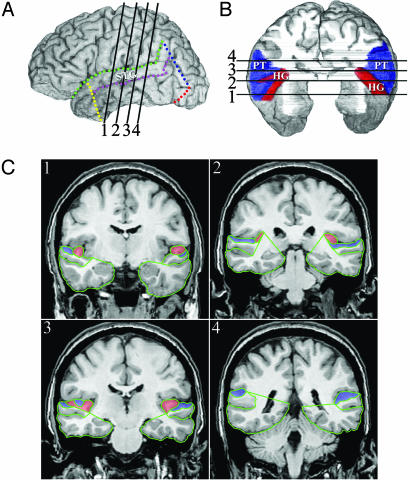
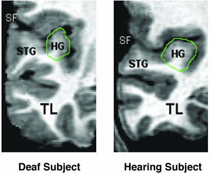
We investigated whether variation in auditory experience in humans during development alters the macroscopic neuroanatomy of primary or auditory association cortices. Volumetric analyses were based on MRI data from 25 congenitally deaf subjects and 25 hearing subjects, all right-handed. The groups were matched for gender and age. Gray and white matter volumes were determined for the temporal lobe, superior temporal gyrus, Heschl's gyrus (HG), and the planum temporale. Deaf and hearing subjects did not differ in the total volume or the gray matter volume of HG, which suggests that auditory deafferentation does not lead to cell loss within primary auditory cortex in humans. However, deaf subjects had significantly larger gray matter-white matter ratios than hearing subjects in HG, with deaf subjects exhibiting significantly less white matter in both left and right HG. Deaf subjects also had higher gray matter-white matter ratios in the rest of the superior temporal gyrus, but this pattern was not observed for the temporal lobe as a whole. These findings suggest that auditory deprivation from birth results in less myelination and/or fewer fibers projecting to and from auditory cortices. Finally, the volumes of planum temporale and HG were significantly larger in the left hemisphere for both groups, suggesting that leftward asymmetries within "auditory" cortices do not arise from experience with auditory processing.
Figures
References
-
- Saada, A. A., Niparko, J. K. & Ryugo, D. K. (1996) Brain Res. 736, 315–328. - PubMed
-
- Tierney, T. S., Russell, F. A. & Moore, D. R. (1997) J. Comp. Neurol. 378, 295–306. - PubMed
-
- Moore, J. K., Niparko, J. K., Miller, M. R. & Linthicum, F. H. (1994) Am. J. Otol. 15, 588–595. - PubMed
-
- Stanton, S. G. & Harrison, R. V. (2000) J. Comp. Neurol. 426, 117–129. - PubMed
-
- Heid, S., Jahn-Siebert, T. K., Klinke, R., Hartmann, R. & Langner, G. (1997) Hear. Res. 110, 191–199. - PubMed
Publication types
MeSH terms
Grants and funding
LinkOut - more resources
Full Text Sources
Medical
