. 2003 Aug 15;31(16):4836-46.
doi: 10.1093/nar/gkg691.

Alterations in the intracellular level of a protein subunit of human RNase P affect processing of tRNA precursors

Affiliations

Alterations in the intracellular level of a protein subunit of human RNase P affect processing of tRNA precursors

Amit Cohen et al. Nucleic Acids Res. .

Abstract

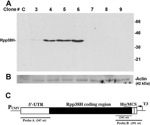
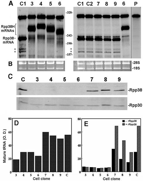
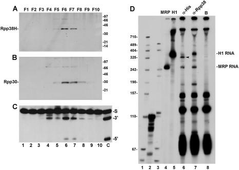
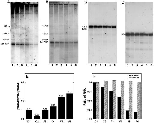
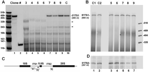
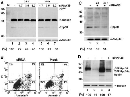
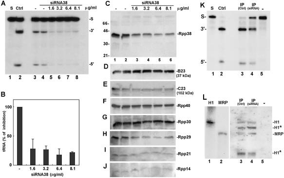
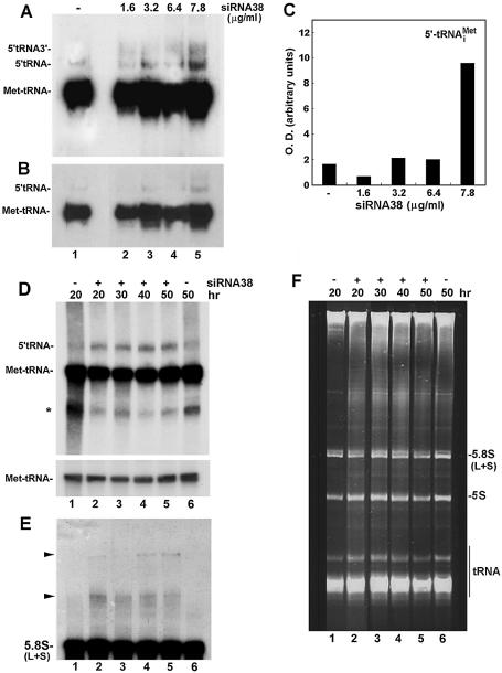
The human ribonucleoprotein ribonuclease P (RNase P), processing tRNA, has at least 10 distinct protein subunits. Many of these subunits, including the autoimmune antigen Rpp38, are shared by RNase MRP, a ribonucleoprotein enzyme required for processing of rRNA. We here show that constitutive expression of exogenous, tagged Rpp38 protein in HeLa cells affects processing of tRNA precursors. Alterations in the site-specific cleavage and in the steady-state level of 3' sequences of the internal transcribed spacer 1 of rRNA are also observed. These processing defects are accompanied by selective shut-off of expression of Rpp38 and by low expression of the tagged protein. RNase P purified from these cells exhibits impaired activity in vitro. Moreover, inhibition of Rpp38 by the use of small interfering RNA causes accumulation of the initiator methionine tRNA precursor. Expression of other protein components, but not of the H1 RNA subunit, is coordinately inhibited. Our results reveal that normal expression of Rpp38 is required for the biosynthesis of intact RNase P and for the normal processing of stable RNA in human cells.


Figures

Figure 1
Figure 1
Constitutive expression of Rpp38H in transfected HeLa S3 cells. (A) S100 crude extracts (100 µg) obtained from eight individual HeLa cell clones transfected with pCI-neo (clone C) or pCIRpp38H (clones 3–9) were subjected to western blot analysis using a monoclonal anti-polyhistidine antibody to test for Rpp38H expression. Proteins were separated by 12% SDS–PAGE, and positions of Rpp38H and protein size markers are indicated. (B) The membrane in (A) was analyzed using a monoclonal antibody against actin, 42 kDa, which serves as an internal control. (C) Schematic map of pCIRpp38H (not drawn to scale) containing a Rpp38 cDNA that has the 5′-UTR and the entire coding region fused to six histidine (His) residues. The cDNA (Rpp38H) is subcloned in the multiple cloning site (MCS) of pCI-neo and transcribed from an upstream cytomegalovirus immediate-early promoter/enhancer (PCMV). Probe A (347 nt) and probe B (291 nt), used for RNase protection analysis, are shown. T3 indicates T3 RNA polymerase used to transcribe probe B.
Figure 2
Figure 2
Shut-off of Rpp38 mRNA and protein expression and impaired RNase P activity in cells expressing Rpp38H. (A) Total RNA extracted from untransfected HeLa cells (clone C1), cells transfected with pCI-neo (clone C2) and cell clones 3–9 (clones 3–9) was subjected to RNase protection analysis using uniformly labeled antisense probe B. Rpp38 mRNA protects 241 nt from the entire 291 nt probe, while Rpp38H mRNAs protect 250–270 nt. Since the probe has sequences derived from the plasmid and a 291 nt RNA was still seen in untransfected cells (clone C1), this RNA represents free probe. The shorter RNA bands indicated by asterisks represent Rpp38 mRNA species. The right-most lane represents undigested probe B (weaker exposure is presented). Numbers between panels indicate single-stranded DNA size markers generated by MspI digestion of pBlueScript. (B) Ethidium bromide staining of 18S and 28S rRNAs, used as internal controls for total RNA analyzed in (A). (C) S100 crude extracts from untransfected cells (clone C) and from cell clones 3–9 were subjected to western blot analysis using affinity purified antibodies against Rpp38 (upper panel) or Rpp30 (lower panel). (D) A representative assay of RNase P activity in S100 crude extracts with equal amounts of protein from clones 3–9 and from untransfected cells using precursor tRNASer as a substrate. Cleavage products were resolved in 8% polyacrylamide/7 M urea and mature tRNA (3′) was quantitated as described in Materials and Methods. (E) Extracts were tested for RNase P activity in the absence (–rRpp38) or presence (+rRpp38) of 8 pmol of highly purified recombinant Rpp38. This recombinant protein has a histidine tag at its N-terminus (5). The optical density values of tRNA are presented in arbitrary units.
Figure 3
Figure 3
Rpp38H is associated with active RNase P that has truncated H1 RNA. (A) S100 crude extracts from cell clone 6 (Fig. 1A) were loaded on a DEAE–Sepharose chromatography column and all the eluted fractions obtained (lanes 1–10) were tested for the presence of Rpp38H as described in Figure 1A. (B) The membrane in (A) was analyzed for Rpp30 using affinity purified antibodies against Rpp30. Proteins were separated by 12% SDS–PAGE, and the positions of Rpp38H, Rpp30 and protein size markers are shown. The protein of Mr ≈ 72 kDa in (A) represents dimers of Rpp38H. (C) RNase P activity in the DEAE eluted fractions as determined by processing of precursor tRNATyr (S) to mature tRNA (3′) and leader sequence (5′). The right-most lane shows a control assay using a DEAE purified RNase P from untransfected cells. The non-specific cleavage of precursor tRNA found in fractions that preceded RNase P activity is commonly seen during fractionation of S100 extracts on a DEAE column (2). (D) 3′ End labeling of RNAs immunoprecipitated with RNase P from whole crude extract of cell clone 6 expressing Rpp38H using anti-polyhistidine antibody (lane 6) or beads alone (lane 8). As a control, anti-Rpp38 antibodies were used to bring down RNase P from normal HeLa cells (lane 7). Positions of the intact 340 nt H1 RNA and 265 nt MRP RNA are shown. The arrow points to truncated H1 RNA. Labeled precursor tRNASer (110 nt) (lane 2) and a MspI digest of pBluescript (lane 1) or pGEM-3 (lane 3) were used as size markers.
Figure 4
Figure 4
Expression of tRNAs and rRNAs in HeLa cells that express Rpp38H. (A) Total RNA extracted from untransfected cells (lane 1), pCI-neo- transfected cells (lane 2) and clones 3–6 expressing Rpp38H (lanes 3–6) was analyzed by northern blot hybridization analysis using a uniformly labeled antisense RNA probe for precursor tRNAiMet. This probe detects the mature tRNAiMet, 75 nt in length, and the precursor tRNAiMet (ptRNAiMet, 95 nt). (BD) The blot in (A) was rehybridized with antisense RNA probes against human precursor tRNASer, the 157 nt 5.8S rRNA (large and small species) or the 121 nt 5S rRNA, respectively. The signal seen at the top of (A) is non-specific hybridization, which was not seen in panels (B)–(D). (E) The ratio of ptRNAiMet to ptRNAiMet + tRNAiMet in each lane of (A) was determined after quantitation of RNA. (F) tRNAiMet, 5S rRNA and 5.8SS rRNA in (A), (C) and (D) were quantitated and the ratios tRNAiMet:5S rRNA and 5.8SS rRNA:5S rRNA are plotted.
Figure 5
Figure 5
Alterations in the processing pattern and steady-state level of 3′ ITS1 rRNA in cells expressing Rpp38H. (A) Total RNA extracted from cell clones 3–6 (lanes 3–6), clones 7–9 (lanes 7–9) and untransfected HeLa cells (lane 10) was subjected to RNase protection analysis using probe P2 (lane 1). This probe protects 488 nt of the 3′ sequence of ITS1 rRNA. The two other protected RNAs of ∼230 and 350 nt seen in control cells (lane 10, small arrows), but not in clones 3–6 (lanes 3–6), represent specific cleavage sites in the 3′ ITS1 rRNA sequence. Lower bands may be generated from partial digestion of the probe as a result of forming intramolecular stem–loop rRNA structures. Labeled precursor tRNATyr (131 nt, lane 2) and MspI-digested pBlueScript were used as size markers. Protected RNAs were separated in 5% polyacrylamide/7 M urea gels. (B) RNA from untransfected cells (lane 1), pCI-neo-transfected cells (lane 2), cell clones 5 and 6 (lanes 3 and 4) and clones 7–9 (lanes 5–7) was analyzed by RNase protection analysis using probe P1 that protects 568 nt of the 5′ sequence of ITS1 rRNA. (C) The antisense RNA probes P1, P2 and P3, which cover different regions in ITS1 and ITS2 that separate 18S, 5.8S and 28S rRNAs, are indicated. The arrow points to a putative cleavage site in the 3′ end of ITS1. (D) Total RNA seen in (B) was analyzed using probe P3 that protects 257 nt of the 3′ end of ITS2 rRNA.
Figure 6
Figure 6
Inhibition of expression of Rpp38 in HeLa cells transfected with siRNA38. (A) Equal amounts of proteins in S20 crude extracts of HeLa cells transfected for 24 (left panel) or 48 h (right panel) with 1.7, 3.5 or 5.2 µg/ml siRNA38 were separated by 12% SDS–PAGE and then subjected to western blot analysis using anti-Rpp38 and anti-γ-tubulin antibodies. Positions of Rpp38, γ-tubulin and protein size markers are shown. Numbers below panels represent percent inhibition of Rpp38, as determined by optical density scans of the band of Rpp38 and normalized to that of γ-tubulin. Percentage inhibition is relative to the control cells. (B) FACS analysis of siRNA38-treated (left) and mock-treated (right) HeLa cells using an Annexin V and propidium iodide (PI) kit (from Roche Molecular Biochemicals, Germany). Percentage of living (lower left quadrant), apoptotic (lower right) and necrotic (upper right) cells are shown. (C) Equal amounts of proteins in S20 crude extracts of 293 HEK cells transfected for 24 (lane 2) or 48 h (lane 3) with 5.2 µg/ml siRNA38 were separated by 12% SDS–PAGE and then subjected to western blot analysis using anti-Rpp38 antibody (upper panel) or anti-γ-tubulin antibody (lower panel). Positions of Rpp38, γ-tubulin and protein size markers are indicated. Numbers below the panel represent percent inhibition of Rpp38, as determined by optical density scans of the band of Rpp38 normalized to that of γ-tubulin. (D) HeLa cells were transfected for 48 h with pEGFP-Rpp38 or pEGFP-Rpp38Δ in the presence (lanes 2 and 4) or absence (lanes 1 and 3) of 5.2 µg/ml siRNA38. Equal amounts of protein in S20 crude extracts of the cells were subjected to western blot analysis as described in (A). Numbers below the panel represent percentage inhibition of the fused proteins.
Figure 7
Figure 7
Inhibition of RNase P activity in extracts of HeLa cells transfected with siRNA38. (A) Whole crude extracts of HeLa cells transfected for 48 h with 1.6, 3.2, 6.4 or 8.1 µg/ml siRNA38 (lanes 5–8) were tested for the activity of RNase P in processing of labeled precursor tRNATyr. Control extracts of cells mock-treated for 24 and 48 h are shown in lanes 3 and 4. Cleavage was for 4 min at 37°C so as to be in the linear range (<30% cleavage of substrate in control extracts). The positions of substrate (S) and cleavage products, the 5′ leader sequence (5′) and tRNA (3′) are indicated. Ctrl represents enzymatic assay of DEAE-purified HeLa RNase P (lane 2). (B) Quantitation of processing of precursor tRNATyr seen in (A). Values are the mean of three independent experiments whose standard deviation is shown by the bars. (CJ) Whole crude extracts of cells seen in (A) were subjected to western blot analyses using antibodies against Rpp38 (C), B23 (D), C23 (E), Rpp40 (F), Rpp30 (G), Rpp29 (H), Rpp21 (I) and Rpp14 (J). Positions of protein subunits and size markers are indicated. (K) Processing of precursor tRNATyr by RNase P immunoprecipitated by anti-Rpp30 antibody from S20 crude extracts of HeLa cells treated with 8.1 µg/ml siRNA38 (lane 4) or left untreated (lane 3). Lane 5 indicates beads not coupled to antibody and lane 3 shows activity of RNase P brought down by anti-Rpp30 antibodies from equal amounts of proteins in S20 crude extracts of untreated HeLa cells. (L) Northern blot analysis of RNA that was extracted from the immunoprecipitates described in (K) (lanes 3–5). Labeled, antisense H1 RNA and MRP RNA were used as probes (see Materials and Methods). The RNA bands indicated by asterisks represent truncated H1 RNA molecules (∼310 and 170 nt), which can also be detected by the use of an antisense H1 RNA alone (; data not shown). As a control, sense H1 and MRP RNAs were analyzed (lanes 1 and 2) and a shorter exposure of the blot (lanes 1 and 2) is attached.
Figure 8
Figure 8
Processing of tRNA and 5.8S rRNA in siRNA38-treated HeLa cells. (A) Total RNA was extracted from HeLa cells treated with siRNA38 and then subjected to northern blot hybridization analysis using an antisense RNA probe against tRNAiMet. 5′-tRNA-3′ represents the primary transcript with 5′ leader sequence and 3′ trailer. 5′-tRNA represents a precursor with 5′ leader sequence. (B) A shorter exposure of the gel seen in (A). (C) Quantitation of the 5′-tRNA seen in (A). (D) Cells were transfected with 7.8 µg/ml siRNA38 for the indicated time points and total RNA was extracted and analyzed as in (A). The asterisk may represent uncharged or truncated tRNA. The lower panel depicts shorter exposure of mature tRNAiMet. (E) The membrane shown in (D) was rehybridized with an antisense oligonucleotide against 5.8S rRNA. Arrows point to large RNA transcripts containing 5.8S rRNA sequences. (F) Ethidium bromide staining of total RNA separated in denaturing 8% polyacrylamide/7 M urea gels. tRNAs, 5S rRNA and small and large 5.8S rRNAs are indicated.

Similar articles

Cited by

References

    1. Altman S., Gopalan,V. and Vioque,A. (2000) Varieties of RNase P: a nomenclature problem? RNA, 6, 1689–1694. - PMC - PubMed
    1. Jarrous N. (2002) Human ribonuclease P: subunits, function and intranuclear localization. RNA, 8, 1–7. - PMC - PubMed
    1. Lygerou Z., Pluk,H., van Venrooij,W.J. and Seraphin,B. (1996) hPop1: an autoantigenic protein subunit shared by the human RNase P and RNase MRP ribonucleoproteins. EMBO J., 15, 5936–5948. - PMC - PubMed
    1. Eder P.S., Kekuda,R., Stolc,V. and Altman,S. (1997) Characterization of two scleroderma autoimmune antigens that copurify with human ribonuclease P. Proc. Natl Acad. Sci. USA, 91, 1101–1106. - PMC - PubMed
    1. Jarrous N., Eder,P.S., Guerrier-Takada,C., Hoog,C. and Altman,S. (1998) Autoantigenic properties of some protein subunits of catalytically active complexes of human ribonuclease P. RNA, 4, 407–417. - PMC - PubMed

Publication types

MeSH terms